Copyright
©2008 The WJG Press and Baishideng.
World J Gastroenterol. Feb 28, 2008; 14(8): 1204-1211
Published online Feb 28, 2008. doi: 10.3748/wjg.14.1204
Published online Feb 28, 2008. doi: 10.3748/wjg.14.1204
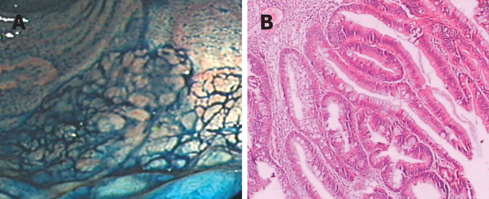
Figure 1 Endoscope and the histopathology of LST.
A: LST under endoscope which grows laterally in the rectum, 3 cm far from the anus and has a diameter of 60 mm × 70 mm; B: The histopathology of the tissue: a villous adenoma accompanied by moderate severe atypical hyperplasia.
- Citation: Wang XY, Lai ZS, Yeung CM, Wang JD, Deng W, Li HY, Han YJ, Kung HF, Jiang B, Lin MCM. Establishment and characterization of a new cell line derived from human colorectal laterally spreading tumor. World J Gastroenterol 2008; 14(8): 1204-1211
- URL: https://www.wjgnet.com/1007-9327/full/v14/i8/1204.htm
- DOI: https://dx.doi.org/10.3748/wjg.14.1204


