Copyright
©2008 The WJG Press and Baishideng.
World J Gastroenterol. Feb 21, 2008; 14(7): 1016-1022
Published online Feb 21, 2008. doi: 10.3748/wjg.14.1016
Published online Feb 21, 2008. doi: 10.3748/wjg.14.1016
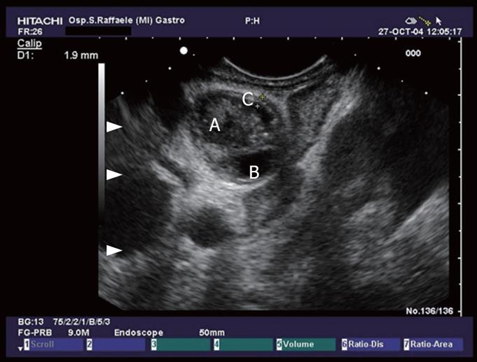
Figure 4 Hepatic ilum seen from the duodenal bulb: common bile duct with sludge (A), thickening of the CBD wall (C), and cystic duct (B).
- Citation: Petrone MC, Arcidiacono PG, Testoni PA. Endoscopic ultrasonography for evaluating patients with recurrent pancreatitis. World J Gastroenterol 2008; 14(7): 1016-1022
- URL: https://www.wjgnet.com/1007-9327/full/v14/i7/1016.htm
- DOI: https://dx.doi.org/10.3748/wjg.14.1016


