Copyright
©2008 The WJG Press and Baishideng.
World J Gastroenterol. Feb 21, 2008; 14(7): 1016-1022
Published online Feb 21, 2008. doi: 10.3748/wjg.14.1016
Published online Feb 21, 2008. doi: 10.3748/wjg.14.1016
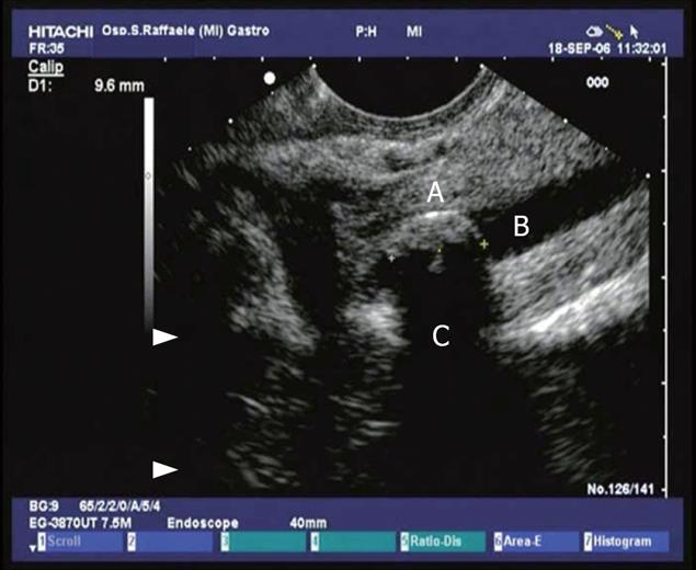
Figure 1 Conventional endosonographic imaging (7.
5 MHz) of choledocholithiasis. A 9.6 mm stone (A) is seen as an hyperechoic structure with acoustic shadowing (C) in the intrapancreatic tract of the common bile duct (B).
- Citation: Petrone MC, Arcidiacono PG, Testoni PA. Endoscopic ultrasonography for evaluating patients with recurrent pancreatitis. World J Gastroenterol 2008; 14(7): 1016-1022
- URL: https://www.wjgnet.com/1007-9327/full/v14/i7/1016.htm
- DOI: https://dx.doi.org/10.3748/wjg.14.1016


