Copyright
©2008 The WJG Press and Baishideng.
World J Gastroenterol. Feb 14, 2008; 14(6): 892-898
Published online Feb 14, 2008. doi: 10.3748/wjg.14.892
Published online Feb 14, 2008. doi: 10.3748/wjg.14.892
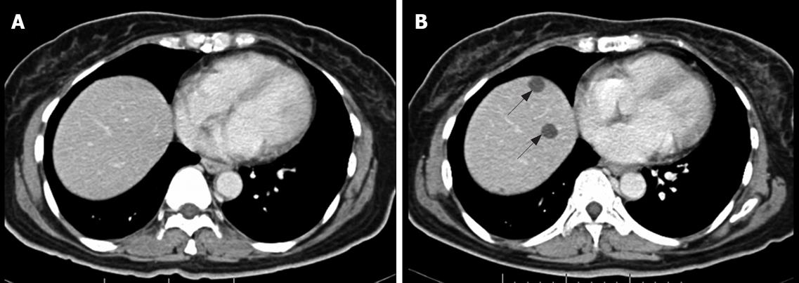
Figure 5 NC pattern.
Contrast-enhanced CT scan of abdomen in 50-year-old woman with metastatic GIST in the liver. A: Before imatinib treatment, no liver metastases were visible at this level; B: Contrast-enhanced CT after 2 mo treatment with imatinib. At least two non-enhancing low-density lesions were newly seen at the dome of the liver (arrows).
- Citation: Phongkitkarun S, Phaisanphrukkun C, Jatchavala J, Sirachainan E. Assessment of gastrointestinal stromal tumors with computed tomography following treatment with imatinib mesylate. World J Gastroenterol 2008; 14(6): 892-898
- URL: https://www.wjgnet.com/1007-9327/full/v14/i6/892.htm
- DOI: https://dx.doi.org/10.3748/wjg.14.892


