Copyright
©2008 The WJG Press and Baishideng.
World J Gastroenterol. Feb 14, 2008; 14(6): 892-898
Published online Feb 14, 2008. doi: 10.3748/wjg.14.892
Published online Feb 14, 2008. doi: 10.3748/wjg.14.892
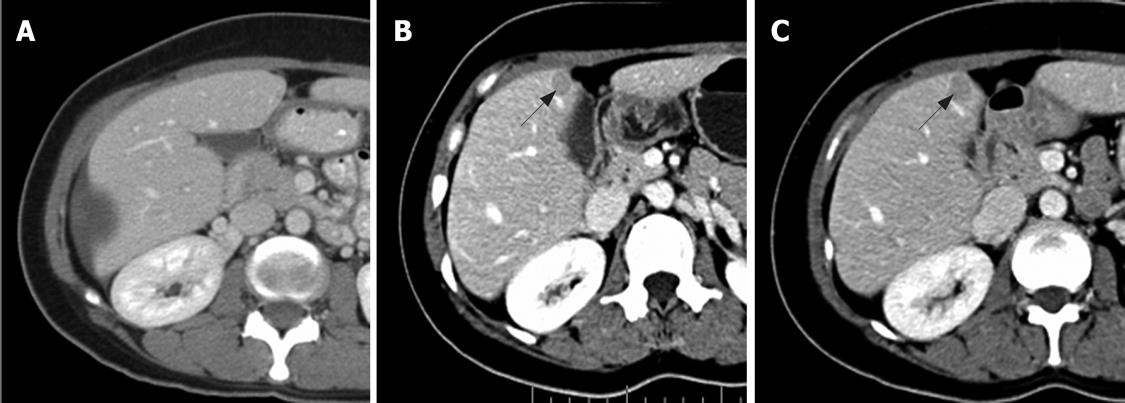
Figure 3 NS pattern.
Contrast-enhanced CT scan of abdomen obtained in a 39-year-old woman with metastatic GIST in the liver. A: Before imatinib treatment, a large subcapsular cystic lesion in the right hepatic lobe and a small amount of free fluid were noted; B: After 10 mo therapy, there were new, well-defined, homogeneous enhancing nodules adjacent to the gallbladder (arrow); C: After 13 mo therapy, there was a slight increase in size of the mentioned nodule (arrow).
- Citation: Phongkitkarun S, Phaisanphrukkun C, Jatchavala J, Sirachainan E. Assessment of gastrointestinal stromal tumors with computed tomography following treatment with imatinib mesylate. World J Gastroenterol 2008; 14(6): 892-898
- URL: https://www.wjgnet.com/1007-9327/full/v14/i6/892.htm
- DOI: https://dx.doi.org/10.3748/wjg.14.892


