Copyright
©2008 The WJG Press and Baishideng.
World J Gastroenterol. Feb 14, 2008; 14(6): 892-898
Published online Feb 14, 2008. doi: 10.3748/wjg.14.892
Published online Feb 14, 2008. doi: 10.3748/wjg.14.892
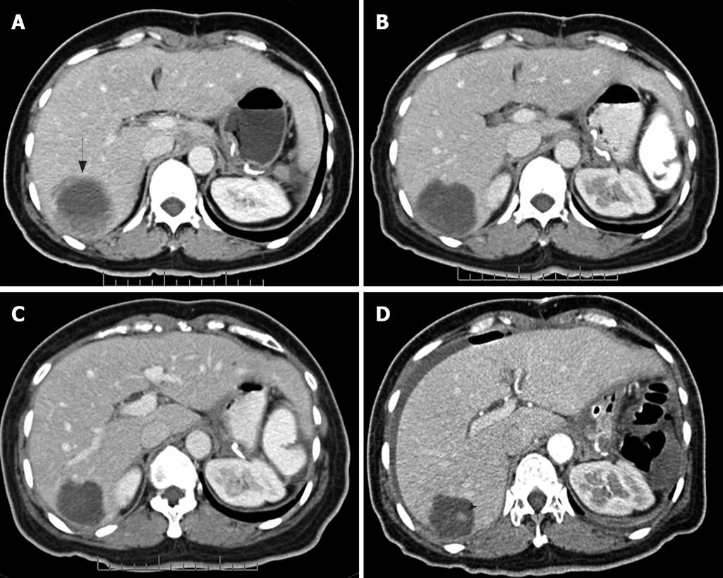
Figure 1 FP pattern.
Contrast-enhanced CT of the abdomen obtained from a 39-year-old woman with metastatic GIST in the right lobe of liver. A: Before imatinib therapy, the lesion showed ill-defined thickened rim enhancement (arrow); B: After 2 mo therapy, a nearly complete cystic change and a thin lesion boundary were observed; C: After 5 mo therapy, a further decrease in size (partial response by RECIST) was seen; D: After 10 mo therapy, a small enhancing nodule was seen. Nodule within a mass or FP (arrow head).
- Citation: Phongkitkarun S, Phaisanphrukkun C, Jatchavala J, Sirachainan E. Assessment of gastrointestinal stromal tumors with computed tomography following treatment with imatinib mesylate. World J Gastroenterol 2008; 14(6): 892-898
- URL: https://www.wjgnet.com/1007-9327/full/v14/i6/892.htm
- DOI: https://dx.doi.org/10.3748/wjg.14.892


