Copyright
©2008 The WJG Press and Baishideng.
World J Gastroenterol. Feb 14, 2008; 14(6): 864-875
Published online Feb 14, 2008. doi: 10.3748/wjg.14.864
Published online Feb 14, 2008. doi: 10.3748/wjg.14.864
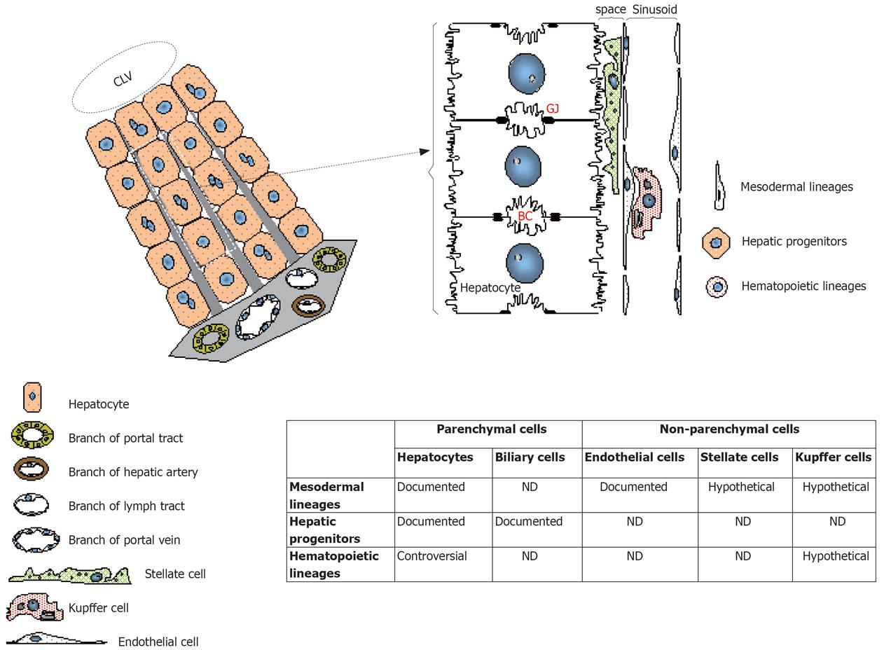
Figure 3 Involvement of stem cells in the regeneration of the liver parenchymal and non-parenchymal cell lineages.
Schema shows the liver architecture and details the topography of the cell populations. In the table are resumed the cell fractions that could potentially be the targets of differentiation after transplantation of stem cells. Some of these differentiation potentials were documented with stem cells while other were contradicted and were thus exposed as controversial. Other differentiation pathways are purely hypothetical based on the embryonic origin of the stem cells. BC: Bile canalicule; CLV: Centrolobular vein; GJ: Gap junctions.
- Citation: Lysy PA, Campard D, Smets F, Najimi M, Sokal EM. Stem cells for liver tissue repair: Current knowledge and perspectives. World J Gastroenterol 2008; 14(6): 864-875
- URL: https://www.wjgnet.com/1007-9327/full/v14/i6/864.htm
- DOI: https://dx.doi.org/10.3748/wjg.14.864


