Copyright
©2008 The WJG Press and Baishideng.
World J Gastroenterol. Feb 14, 2008; 14(6): 853-863
Published online Feb 14, 2008. doi: 10.3748/wjg.14.853
Published online Feb 14, 2008. doi: 10.3748/wjg.14.853
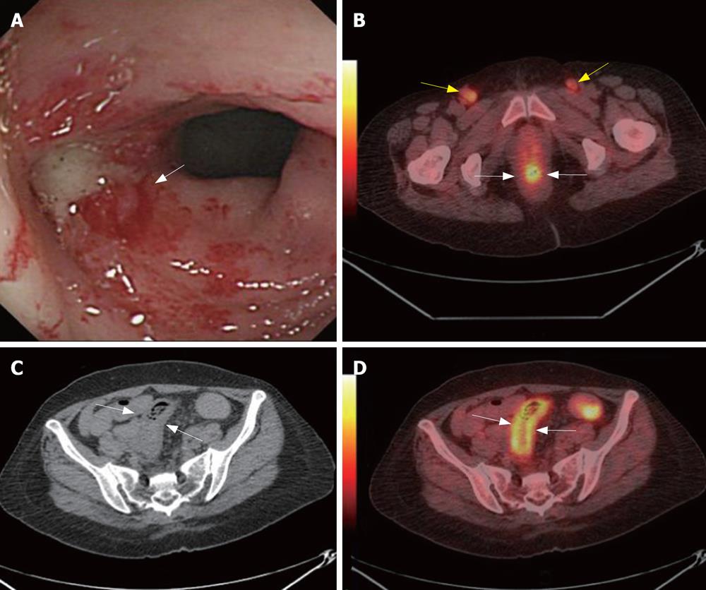
Figure 1 A 56-year-old woman was suffered from uterine cervix cancer and accepted local radiation and chemotherapy 2-mo ago.
CC found enterocolitis of sigmoid colon (white arrow, A). PET/CT detected high metabolism metastasis lymphoid nodes at both sides of the inguina (yellow arrows, B) and rectum wall was high metabolism (white arrows, B). CT showed thickening of sigmoid colon wall (white arrows, C). PET/CT illustrated sigmoid colon wall was high metabolism (white arrows, D). Biopsy followed by PET/CT revealed metastasis lymphoid nodes at both sides of the inguina. The case illustrated the potential value of PET/CTC in radiation enterocolitis.
- Citation: Sun L, Wu H, Guan YS. Colonography by CT, MRI and PET/CT combined with conventional colonoscopy in colorectal cancer screening and staging. World J Gastroenterol 2008; 14(6): 853-863
- URL: https://www.wjgnet.com/1007-9327/full/v14/i6/853.htm
- DOI: https://dx.doi.org/10.3748/wjg.14.853


