Copyright
©2008 The WJG Press and Baishideng.
World J Gastroenterol. Feb 7, 2008; 14(5): 764-770
Published online Feb 7, 2008. doi: 10.3748/wjg.14.764
Published online Feb 7, 2008. doi: 10.3748/wjg.14.764
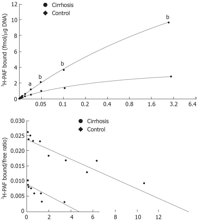
Figure 2 A: Saturation curve binding to cirrhotic and normal rat Kupffer cells.
Cells were used after overnight culture for receptor binding assay as described in the Method section. 3H-PAF at a concentration between 0.125 and 3.2 nmol/L was incubated with Kupffer cells in presence or absence of 5 &mgr;mol/L unlabeled PAF at 22°C for 3 h. aP < 0.05, bP < 0.01 vs control; B: Scattered blot analysis of 3H-PAF binding to cirrhotic and normal rat Kupffer cells. The results were analyzed by scatchard plot. Cirrhosis: R = 0.99, Kd = 2.08 nmol/L, Bax = 27.1882 ± 2.0885 fmol/mg. DNA; Control: R = 0.96, Kd = 1.57 nmol/L, Bax = 4.4024 ± 0.3155 fmol/mg. DNA.
- Citation: Lu YY, Wang CP, Zhou L, Chen Y, Su SH, Feng YY, Yang YP. Synthesis of platelet-activating factor and its receptor expression in Kupffer cells in rat carbon tetrachloride-induced cirrhosis. World J Gastroenterol 2008; 14(5): 764-770
- URL: https://www.wjgnet.com/1007-9327/full/v14/i5/764.htm
- DOI: https://dx.doi.org/10.3748/wjg.14.764


