Rapid Communication Open Access
Copyright ©2008 The WJG Press and Baishideng. All rights reserved.
World J Gastroenterol. Feb 7, 2008; 14(5): 758-763
Published online Feb 7, 2008. doi: 10.3748/wjg.14.758
LINE-1 family member GCRG123 gene is up-regulated in human gastric signet-ring cell carcinoma
Gang-Shi Wang, Meng-Wei Wang, Ben-Yan Wu, Xin-Yan Yang, Wei-Hua Wang, Wei-Di You, Department of Gerontal Gastroenterology, General Hospital of Chinese PLA, Beijing 100853, China
Correspondence to: Dr. Gang-Shi Wang, Department of Gerontal Gastroenterology, General Hospital of Chinese PLA, Beijing 100853, China. wanggangshi@hotmail.com
Telephone: +86-10-66937393
Fax: +86-10-66937393
Received: August 2, 2007
Revised: October 9, 2007
Published online: February 7, 2008

Abstract

AIM: To analyze the expression profiles of a human gastric-cancer-related gene, GCRG123, in human gastric signet-ring cell carcinoma tissues, and to perform bioinformatics analysis on GCRG123.

METHODS: In situ hybridization was used to explore the GCRG123 expression pattern in paraffin-embedded gastric tissues, including 15 cases of signet-ring cell carcinoma, 15 of intestinal-type adenocarcinoma, and 15 of normal gastric mucosa. Northern blotting was used to analyze the differences in GCRG123 expression between stomach signet-ring cell carcinoma and intestinal-type adenocarcinoma tissues. Online software, including BLAST, Multalin and BLAT, were applied for bioinformatics analysis. National Center for Biotechnology Information (NCBI) and the University of California Santa Cruz (UCSC) databases were used for the analyses.

RESULTS: The in situ hybridization signal appeared as blue precipitates restricted to the cytoplasm. Ten out of 15 cases of gastric signet ring cell carcinoma, normal gastric mucosal epithelium and pyloric glands showed high GCRG123 expression. Low GCRG123 expression was observed in gastric intestinal-type adenocarcinoma and normal gastric glands. Northern blotting revealed that GCRG123 was up-regulated in signet-ring cell carcinoma tissue but down-regulated in intestinal-type adenocarcinoma tissue. BLAST and Multalin analyses revealed that the GCRG123 sequence had 92% similarity with the ORF2 sequence of human long interspersed nuclear element retrotransposons (LINE-1, L1). BLAT analysis indicated that GCRG123 mapped to all chromosomes. GCRG123 was found to integrate in the intron-17 and -23 of Rb, 5’ flanking region of IL-2 and clotting factor IX genes.

CONCLUSION: GCRG123, an active member of the L1 family, was up-regulated in human gastric signet-ring cell carcinoma.

Key Words: Stomach, Signet-ring cell carcinoma, GCRG123, LINE-1, In situ hybridization, Bioinformatics


INTRODUCTION

Gastric cancer remains one of the most common forms of cancer worldwide with approximately 870 000 new cases and 650 000 deaths per year[14], which accounts for about 9.9% of new cancers[5]. Worldwide, there has been a decline in the incidence of the intestinal type gastric cancer in the last few decades, following the overall decline in the incidence of gastric cancer. By contrast, the decline in the diffuse type gastric cancer has been more gradual. Some studies have reported an increase in the diffuse type of gastric carcinoma, especially the signet-ring cell type[6]. As a result, the diffuse type now accounts for about 30% of gastric carcinomas in some reported series[7]. Intestinal-type and diffuse-type gastric cancer differ in their epidemiology, pathogenesis, genetic profile, and clinical outcome[8].

In 1999, we cloned a gene segment GCRG123, which was down-regulated in gastric adenocarcinoma. BLASTX analysis revealed that the predicted GCRG123 product was a lamin-like protein[9]. Recently, we have used the updated GeneBank database for further analysis on the GCRG123 sequence. We found that GCRG123 appeared to be a long interspersed nucleotide element-1 (LINE-1, L1) family member. Additionally, we revealed that GCRG123 was ultimately up-regulated in stomach signet-ring cell carcinoma, as well as in normal pyloric glands and epithelia, which shows an opposite expression pattern compared with its down-regulation in stomach intestinal-type adenocarcinoma.

MATERIALS AND METHODS
Patients and tissue acquisition

All samples were obtained from the Department of Pathology, General Hospital of Chinese PLA. Specimens of paraffin-embedded gastric tissues, including 15 cases of gastric signet-ring cell carcinoma, 15 of advanced gastric intestinal-type adenocarcinoma and 15 of normal gastric mucosal tissues, were collected for in situ hybridization analysis. One set of fresh gastric signet-ring cell carcinomas and paired non-cancerous gastric tissues, and one set of fresh gastric intestinal-type adenocarcinomas and paired non-cancerous gastric tissues were used for Northern blot analysis. The diagnosis of cancer was confirmed through histology.

In situ hybridization

cRNA probe labeling: Digoxigenin-labeled anti-sense and sense cRNA probes were prepared by in vitro transcription (DIG RNA Labeling Kit (SP6/T7); Roche Diagnostics, Mannheim, Germany). Briefly, the following labeling procedure was employed: purified GCRG123 cDNA 100 ng/10 &mgr;L, 5 × NTP labeling mixture 4 &mgr;L, 5 × transcription buffer 4 &mgr;L, and RNA polymerase SP6/T7 2 &mgr;L were mixed gently, centrifuged, and then incubated for 1 h at 42°C. Two microliters of RNase-free DNase I was added to remove template DNA, by incubating for 15 min at 37°C, and the reaction was finally stopped by adding 2 &mgr;L 0.2 mol/L EDTA (pH 8.0). Labeling efficiency was directly detected by a spot test as described in the protocol of the kit.

Hybridization: All specimens were fixed in 10% neutral-buffered formalin and embedded in paraffin. A series of 5-&mgr;m thick sections were cut for analysis. In situ hybridization was performed as previously described[1011] with some amendments, using digoxigenin-labeled anti-sense cRNA probes. Briefly, the slides were dried at 40°C overnight, dewaxed, rehydrated and pretreated with DEPC-treated PBS containing 100 mmol/L glycine and 0.3% Triton X-100, respectively. The sections were then permeabilized with 20 &mgr;g/mL RNase-free proteinase K (Merck, Darmstadt, Germany) for 20 min, incubated at 37°C for at least 20 min with prehybridization buffer. Each section was overlaid with 30 &mgr;L hybridization buffer containing 10 ng digoxigenin-labeled cRNA probe and incubated at 42°C overnight. After hybridization, the section was incubated with digoxigenin antibody (75 mU/mL) for 2 h. The positive signal for GCRG123 mRNA was detected by using NBT/BCIP (Promega, WI, USA) as a substrate. Sense cRNA probes were used as a negative control. The presentation of blue staining in the cytoplasm was considered positive. The positive staining of cytoplasm was scored manually as described below: -, barely detectable light blue; 1+, diffuse light blue; 2+, blue; 3+, dark blue. More than 100 non-tumor or tumor cells were quantified in each measurement, and more than one measurement was required to confirm the diagnosis. Consequent slides with H&E staining were then reviewed to compare the histological patterns to the staining patterns in the in situ hybridization preparations.

Northern blot analysis

Dig Northern Starter Kit (Roche Diagnostic, Indianapolis, IN, USA) was used. The procedure of hybridization was performed according to the manufacturer’s protocol. Digoxigenin-labeled cRNA probe was generated as described above. Digoxigenin-labeled sense cRNA probe was used as a negative control. The hybridization signals were visualized with chemiluminescence, which was recorded on X-ray films. The exposure time was 10 min.

Bioinformatic analysis

BLAST (http://www.ncbi.nlm.nih.gov/BLAST/)[12] was used for sequence similarity search. MultAlin (http://bioinfo.genopole-toulouse.prd.fr/multalin/multalin.html)[13] was used for multiple sequence alignment analysis. BLAT (http://genome.ucsc.edu/cgi-bin/hgBlat) was applied to find the genomic coordinates of mRNA, and to search for gene family members[14]. Alignment parameters were set as default. NCBI and UCSC databases were used for the analyses.

Informed consent

The study protocol was approved by the Institutional Review Board of the hospital under the guidelines of the 1975 Declaration of Helsinki. Written informed consent was obtained from all patients.

RESULTS
In situ hybridization

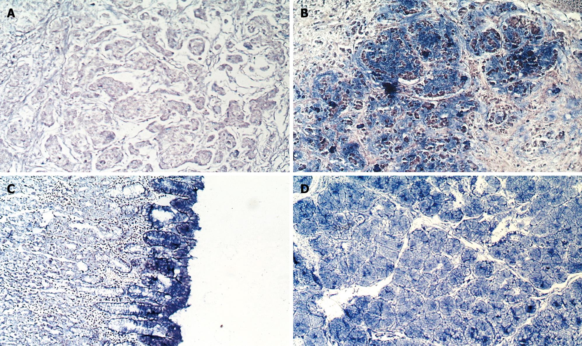
GCRG123 expression was analyzed at the mRNA level using in situ hybridization. The hybridization signal appeared as blue staining in the cytoplasm. Fourteen gastric intestinal-type adenocarcinoma cases showed - to ± staining (Figure 1A). Interestingly, 10 out of 15 gastric signet-ring cell carcinoma tissues were stained 2+ to 3+ (Figure 1B), while the other signet-ring cell tissues were stained ± to +. In the normal gastric mucosa, all gastric epithelia and pyloric glands were heavily stained (grade 2+ to 3+) (Figure 1C and D), however, the normal gastric glands were stained lightly (Figure 1C).

Figure 1
Figure 1 In situ hybridization analysis of GCRG123 in gastric mucosa. GCRG123 showed low expression (blue precipitates restricted to the cytoplasm) in intestinal-type adenocarcinoma (A) and normal gastric glands (C, left region); high expression in signet-ring cell carcinoma (B); normal epithelia (C); and normal pyloric glands (D). NBT/BCIP was used as an alkaline phosphatase substrate (magnification was 10 × 10).
Northern blotting

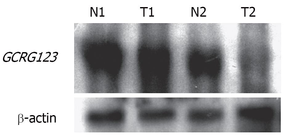
Both normal gastric tissue and signet-ring cell carcinoma tissue showed strong hybridization signals, while the gastric intestinal-type carcinoma tissue showed a weak signal. These results seem to indicate that GCRG123 expression was higher in gastric signet-ring cell carcinoma than in intestinal-type adenocarcinoma (Figure 2).

Figure 2
Figure 2 Northern blotting of GCRG123 in gastric tissues. The expression of β-actin served as an internal control. Total RNA, 1 &mgr;g per lane, was separated on a 1% agarose/formaldehyde gel. After blotting onto a nylon membrane, the membrane was probed with digoxigenin-labeled anti-sense GCRG123 cRNA. N, normal gastric tissue; T, tumor; 1, signet-ring cell carcinoma; 2, intestinal-type adenocarcinoma.
Bioinformatic analysis

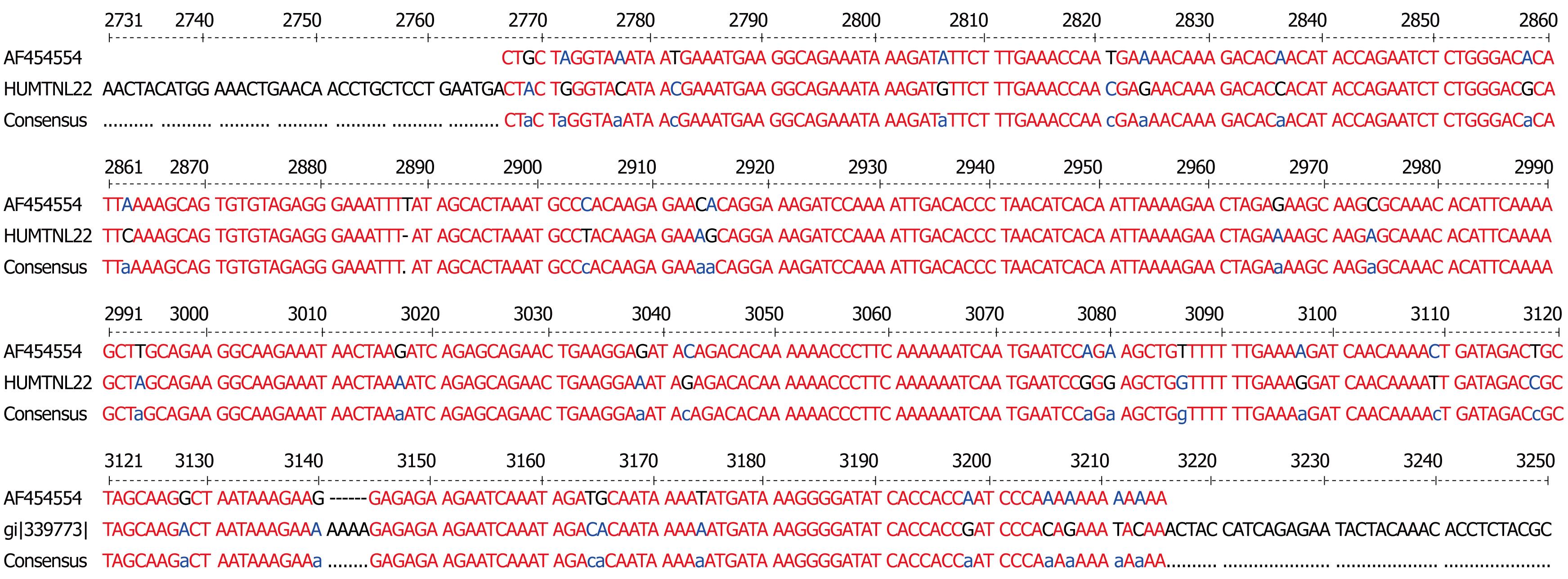
The sequence of GCRG123 was enrolled for a sequence-similarity search in the GenBank database. GCRG123 shared 92% similarity with the human transposon L1 (M80343) sequence, as well as the highly active L1 sequence reported in the literature[15]. Multalin analysis revealed that GCRG123 was located at nucleotides 2767-3215 of L1, at which the L1 ORF2 dwells (Figure 3). GCRG123 was found to integrate in intron-17 and -23 of Rb (L11910), the 5’ flanking region of IL-2 (X67285), and the 5’ flanking region of clotting factor IX (X75349) genes (GenBank accession numbers in parentheses). BLAT analysis showed that GCRG123 mapped to all the human chromosomes.

Figure 3
Figure 3 Multalin analysis of GCRG123 (AF454554) and L1 (HUMTNL22). Red font represents high consensus, blue or black font represents low consensus.
DISCUSSION

Our previous bioinformatic analysis of GCRG123 showed that the predicted peptide sequence of GCRG123 has 80% homology with a human lamin-like protein (GenBank accession number, AAA36178)[9], therefore, we predicted that GCRG123 might be a gene that encoded a lamin-like protein. However, the results of the present study indicate that GCRG123 tends to be an active LINE-1 family member.

Retrotransposable elements, such as L1, Alu and endogenous retroviruses, make up some 45% of human DNA[16]. L1 is one of the most successful retrotransposons and occupies 17% of DNA[17]. In the early stages, because of the lack of any obvious correlation with cellular functions, retrotransposable elements were considered to be mere remnants of our genetic evolution and given the name ‘‘junk DNA’’[18]. However, recent data have revealed that some L1s do remain retrotransposition-competent. It has been shown that L1 is a functionally capable gene. For example, human embryonic stem cells can express endogenous L1 elements[19]. L1 can retrotranspose in neuronal precursors, and can consequently alter the expression of neuronal genes, which in turn, can influence neuronal cell fate in vitro[20]. L1 retrotransposition can also occur in non-dividing somatic cells[21].

A retrotransposition-competent, functional L1 element covers about 6.1 kb and contains a 5’ untranslated region, a 1-kb ORF1 that encodes a protein (p40) with RNA-binding ability, followed by a 3.8-kb ORF2 that codes for a protein (p150) with endonuclease and reverse transcriptase (RT) activities, and a cysteine-histidine-rich domain. The 3’ end of L1 is terminated by a short 3’ untranslated region and a poly(A) tail[2226]. L1 likely integrates into the genomic DNA by a process called target primed reverse transcription and shapes mammalian genomes through many different ways, such as retrotransposition, transduction and gene expression alteration[27]. Although the overwhelming majority of L1s (about 99.8%) are inactive because of 5’ truncations, internal rearrangements and mutations, 80-100 retrotransposition-competent L1s have been predicted in humans. Remarkably, 84% of the retrotransposition capability of these elements has been shown to be present in six highly active L1s[15].

It has been proposed that retrotransposons modulate the expression of specific genes through a transcriptional interference-based mechanism. To exert such effect on neighboring genes, retrotransposons need not necessarily be full-length, nor retrotransposition-competent, but simply endowed with functional promoter elements[28]. Previous studies have shown that some non-full length L1 elements (even shorter than 500 bp) may yield L1 products such as L1 RNA or active RT[2931]. L1 RNAs are also involved in extensive RNA splicing that can radically alter the diversity of expressed RNA forms from these elements, as well as influence their impact on gene expression upon genomic insertion[32].

The state-of-the-art understanding of L1 described above prompted us to propose that GCRG123 may be an active L1 family member, as supported by our findings. First, the GCRG123 sequence shared 92% homology with the conserved sequence of the highly active L1s (hot L1s). Second, GCRG123 can map to all chromosomes, which is similar to L1s. Third, GCRG123 can integrate in the intron or 5’ flanking region of some known genes, which suggests that it has the potential to adjust the function of these genes. Finally, in situ hybridization and Northern blot analysis at the mRNA level showed a distinct GCRG123 expression pattern in different kinds of gastric mucosa cells.

A lot of evidence has indicated that L1 is involved in a wide spectrum of diseases, including cancer[283334]. In colon cancer, the APC gene can be disrupted by somatic insertion of an L1 sequence into the last exon of the gene[35]. Takai et al have found that hypomethylation of L1 is detected in human hepatocellular carcinoma, but never in the surrounding liver tissues, whether or not liver cirrhosis is present[36]. Hypomethylation of L1 has also been revealed in urothelial carcinoma and prostate adenocarcinoma[3739]. In malignant cells, there is a direct correlation between the hypomethylation of 5’ L1 sequences and the presence of L1 proteins, which suggests that elements with hypomethylated 5’ ends are transcriptionally active[40]. The hypomethylation of repetitive elements in cancer is directly linked to the neoplastic process and not a simple consequence of loss of growth control[41]. Previous studies have shown that L1 is essential for the proliferation of tumor cells. The functional knock-out of LINE-1 expression by RNAi reduces the proliferation and promotes the differentiation of tumorigenic human cells[2942]. Up till now, no data about the relationship between L1 and stomach cancer have been reported. The present study on the distinct expression pattern of GCRG123 in gastric signet-ring cell carcinoma may be the first to link an L1 family member to stomach oncogenesis.

During previous decades, several histological classification systems, such as those of Lauren, Nakamura and Ming, have been widely used for gastric adenocarcinoma. These systems agree that the intestinal/differentiated/expending type and diffuse/undifferentiated/infiltrative type differ greatly in terms of epidemiology, pathogenesis, genetic profile, and clinical outcome[84346]. Signet-ring cell carcinoma belongs to the diffuse/undifferentiated/infiltrative type group. Nakamura has indicated that the morphological findings of undifferentiated type carcinoma under light and electron microscopy are similar to both the mucous cells of pyloric crypts and pyloric gland cells[44]. Interestingly, our study found that GCRG123 was over-expressed in pyloric gland cells, epithelia (surface mucous cells) and signet-ring cells, which correlated with Nakagawa’s description of undifferentiated carcinoma. The distinct GCRG123 expression patterns in intestinal-type adenocarcinoma and signet-ring cell carcinoma also support the concept that these two types of carcinoma have different genetic profiles. GCRG123 is over-expressed simultaneously in gastric epithelia, pyloric glands and signet-ring cell carcinoma, therefore, we believe that GCRG123 might be closely associated with gastric mucous secretion. Further studies are required to confirm this speculation.

COMMENTS
Background

Evidence indicates that a retrotransposition-competent, functional L1 element is involved in a wide spectrum of diseases, including cancer. A previous study has shown that a gene segment GCRG123 is down-regulated in human gastric intestinal-type adenocarcinoma. Intestinal- and diffuse-type gastric cancers differ in their epidemiology, pathogenesis, genetic profile and clinical outcome.

Research frontiers

To modulate the expression of specific genes, retrotransposons, such as L1, need not necessarily be full-length, nor retrotransposition-competent, but simply endowed with functional promoter elements. Some non-full length L1 elements could yield L1 products such as L1 RNA or active RT. L1 is essential for the proliferation of tumor cells. The functional knock-out of L1 expression reduces cell proliferation and promotes cell differentiation.

Innovations and breakthroughs

GCRG123 may be an active L1 family member, because of its sequence homology with the conserved sequence of the highly active L1s and its integration in the intron or 5’ flanking region of some known genes. In situ hybridization and Northern blot analysis showed that GCRG123 was up-regulated in signet-ring cell carcinoma. Besides, GCRG123 was also over-expressed in normal gastric epithelia and pyloric glands. The present study possibly represents the first to link an L1 family member to stomach oncogenesis.

Applications

Updated knowledge about L1 shifts the focus from protein-coding genes to repeated retroelements as mediators of abnormal biological processes. Functional L1 family members may be regarded as promising targets in the development of novel, differentiation-inducing approaches to cancer therapy. The distinct expression pattern of GCRG123 in gastric signet-ring cell carcinoma may provide a new area to further understand the genetic characteristics of gastric signet-ring cell carcinoma.

Terminology

LINE-1: highly repeated sequences, 6-8 K base pairs in length, which contain RNA polymerase II promoters. They also have an ORF that is related to the RT of retroviruses, but they do not contain long terminal repeats. Copies of the LINE 1 (L1) family form about 17% of the human genome.

Peer review

This is an interesting article. The title reflects the major contents of the article. The abstract gives a clear delineation of the research background, objectives, materials and methods, results and conclusions. The methods are innovative and systematic. A detailed description has been provided. The results provide sufficient experimental evidence or data from which scientific conclusions can be drawn. The discussion is well organized and an overall theoretical analysis is given. The most current literature is cited. Overall, a it is well-written and well-researched article.

Footnotes

Supported by the Key Project from the 10th Five-year Plan of Chinese PLA, No. 01Z035, and International Cooperation Project from the 11th Five-year Plan of Chinese PLA, No. 06H044

References
1.  Parkin DM, Bray F, Ferlay J, Pisani P. Global cancer statistics, 2002. CA Cancer J Clin. 2005;55:74-108.  [PubMed]  [DOI]  [Cited in This Article: ]
2.  Lau M, Le A, El-Serag HB. Noncardia gastric adenocarcinoma remains an important and deadly cancer in the United States: secular trends in incidence and survival. Am J Gastroenterol. 2006;101:2485-2492.  [PubMed]  [DOI]  [Cited in This Article: ]
3.  Crew KD, Neugut AI. Epidemiology of gastric cancer. World J Gastroenterol. 2006;12:354-362.  [PubMed]  [DOI]  [Cited in This Article: ]
4.  Kelley JR, Duggan JM. Gastric cancer epidemiology and risk factors. J Clin Epidemiol. 2003;56:1-9.  [PubMed]  [DOI]  [Cited in This Article: ]
5.  Parkin DM. Epidemiology of cancer: global patterns and trends. Toxicol Lett. 1998;102-103:227-234.  [PubMed]  [DOI]  [Cited in This Article: ]
6.  Henson DE, Dittus C, Younes M, Nguyen H, Albores-Saavedra J. Differential trends in the intestinal and diffuse types of gastric carcinoma in the United States, 1973-2000: increase in the signet ring cell type. Arch Pathol Lab Med. 2004;128:765-770.  [PubMed]  [DOI]  [Cited in This Article: ]
7.  Ikeda Y, Mori M, Kamakura T, Haraguchi Y, Saku M, Sugimachi K. Improvements in diagnosis have changed the incidence of histological types in advanced gastric cancer. Br J Cancer. 1995;72:424-426.  [PubMed]  [DOI]  [Cited in This Article: ]
8.  Hamilton JP, Meltzer SJ. A review of the genomics of gastric cancer. Clin Gastroenterol Hepatol. 2006;4:416-425.  [PubMed]  [DOI]  [Cited in This Article: ]
9.  Wang G, Wang M, Wu B, You W. A lamin-like protein gene is down-regulated in human gastric cancer. Zhonghua Yixue Yichuanxue Zazhi. 2003;20:119-122.  [PubMed]  [DOI]  [Cited in This Article: ]
10.  Komminoth P. Digoxigenin as an alternative probe labeling for in situ hybridization. Diagn Mol Pathol. 1992;1:142-150.  [PubMed]  [DOI]  [Cited in This Article: ]
11.  Komminoth P, Merk FB, Leav I, Wolfe HJ, Roth J. Comparison of 35S- and digoxigenin-labeled RNA and oligonucleotide probes for in situ hybridization. Expression of mRNA of the seminal vesicle secretion protein II and androgen receptor genes in the rat prostate. Histochemistry. 1992;98:217-228.  [PubMed]  [DOI]  [Cited in This Article: ]
12.  Altschul SF, Gish W, Miller W, Myers EW, Lipman DJ. Basic local alignment search tool. J Mol Biol. 1990;215:403-410.  [PubMed]  [DOI]  [Cited in This Article: ]
13.  Corpet F. Multiple sequence alignment with hierarchical clustering. Nucleic Acids Res. 1988;16:10881-10890.  [PubMed]  [DOI]  [Cited in This Article: ]
14.  Kent WJ. BLAT--the BLAST-like alignment tool. Genome Res. 2002;12:656-664.  [PubMed]  [DOI]  [Cited in This Article: ]
15.  Brouha B, Schustak J, Badge RM, Lutz-Prigge S, Farley AH, Moran JV, Kazazian HH Jr. Hot L1s account for the bulk of retrotransposition in the human population. Proc Natl Acad Sci USA. 2003;100:5280-5285.  [PubMed]  [DOI]  [Cited in This Article: ]
16.  Deininger PL, Moran JV, Batzer MA, Kazazian HH Jr. Mobile elements and mammalian genome evolution. Curr Opin Genet Dev. 2003;13:651-658.  [PubMed]  [DOI]  [Cited in This Article: ]
17.  Lander ES, Linton LM, Birren B, Nusbaum C, Zody MC, Baldwin J, Devon K, Dewar K, Doyle M, FitzHugh W. Initial sequencing and analysis of the human genome. Nature. 2001;409:860-921.  [PubMed]  [DOI]  [Cited in This Article: ]
18.  Orgel LE, Crick FH. Selfish DNA: the ultimate parasite. Nature. 1980;284:604-607.  [PubMed]  [DOI]  [Cited in This Article: ]
19.  Garcia-Perez JL, Marchetto MC, Muotri AR, Coufal NG, Gage FH, O'Shea KS, Moran JV. LINE-1 retrotransposition in human embryonic stem cells. Hum Mol Genet. 2007;16:1569-1577.  [PubMed]  [DOI]  [Cited in This Article: ]
20.  Muotri AR, Chu VT, Marchetto MC, Deng W, Moran JV, Gage FH. Somatic mosaicism in neuronal precursor cells mediated by L1 retrotransposition. Nature. 2005;435:903-910.  [PubMed]  [DOI]  [Cited in This Article: ]
21.  Kubo S, Seleme MC, Soifer HS, Perez JL, Moran JV, Kazazian HH Jr, Kasahara N. L1 retrotransposition in nondividing and primary human somatic cells. Proc Natl Acad Sci USA. 2006;103:8036-8041.  [PubMed]  [DOI]  [Cited in This Article: ]
22.  McMillan JP, Singer MF. Translation of the human LINE-1 element, L1Hs. Proc Natl Acad Sci USA. 1993;90:11533-11537.  [PubMed]  [DOI]  [Cited in This Article: ]
23.  Singer MF, Krek V, McMillan JP, Swergold GD, Thayer RE. LINE-1: a human transposable element. Gene. 1993;135:183-188.  [PubMed]  [DOI]  [Cited in This Article: ]
24.  Moran JV, Holmes SE, Naas TP, DeBerardinis RJ, Boeke JD, Kazazian HH Jr. High frequency retrotransposition in cultured mammalian cells. Cell. 1996;87:917-927.  [PubMed]  [DOI]  [Cited in This Article: ]
25.  Feng Q, Moran JV, Kazazian HH Jr, Boeke JD. Human L1 retrotransposon encodes a conserved endonuclease required for retrotransposition. Cell. 1996;87:905-916.  [PubMed]  [DOI]  [Cited in This Article: ]
26.  Hohjoh H, Singer MF. Sequence-specific single-strand RNA binding protein encoded by the human LINE-1 retrotransposon. EMBO J. 1997;16:6034-6043.  [PubMed]  [DOI]  [Cited in This Article: ]
27.  Ostertag EM, Kazazian HH Jr. Biology of mammalian L1 retrotransposons. Annu Rev Genet. 2001;35:501-538.  [PubMed]  [DOI]  [Cited in This Article: ]
28.  Sinibaldi-Vallebona P, Lavia P, Garaci E, Spadafora C. A role for endogenous reverse transcriptase in tumorigenesis and as a target in differentiating cancer therapy. Genes Chromosomes Cancer. 2006;45:1-10.  [PubMed]  [DOI]  [Cited in This Article: ]
29.  Kuo KW, Sheu HM, Huang YS, Leung WC. Expression of transposon LINE-1 is relatively human-specific and function of the transcripts may be proliferation-essential. Biochem Biophys Res Commun. 1998;253:566-570.  [PubMed]  [DOI]  [Cited in This Article: ]
30.  Boissinot S, Entezam A, Furano AV. Selection against deleterious LINE-1-containing loci in the human lineage. Mol Biol Evol. 2001;18:926-935.  [PubMed]  [DOI]  [Cited in This Article: ]
31.  Liu J, Nau MM, Zucman-Rossi J, Powell JI, Allegra CJ, Wright JJ. LINE-I element insertion at the t(11;22) translocation breakpoint of a desmoplastic small round cell tumor. Genes Chromosomes Cancer. 1997;18:232-239.  [PubMed]  [DOI]  [Cited in This Article: ]
32.  Belancio VP, Hedges DJ, Deininger P. LINE-1 RNA splicing and influences on mammalian gene expression. Nucleic Acids Res. 2006;34:1512-1521.  [PubMed]  [DOI]  [Cited in This Article: ]
33.  Schulz WA, Steinhoff C, Florl AR. Methylation of endogenous human retroelements in health and disease. Curr Top Microbiol Immunol. 2006;310:211-250.  [PubMed]  [DOI]  [Cited in This Article: ]
34.  Druker R, Whitelaw E. Retrotransposon-derived elements in the mammalian genome: a potential source of disease. J Inherit Metab Dis. 2004;27:319-330.  [PubMed]  [DOI]  [Cited in This Article: ]
35.  Miki Y, Nishisho I, Horii A, Miyoshi Y, Utsunomiya J, Kinzler KW, Vogelstein B, Nakamura Y. Disruption of the APC gene by a retrotransposal insertion of L1 sequence in a colon cancer. Cancer Res. 1992;52:643-645.  [PubMed]  [DOI]  [Cited in This Article: ]
36.  Takai D, Yagi Y, Habib N, Sugimura T, Ushijima T. Hypome-thylation of LINE1 retrotransposon in human hepatocellular carcinomas, but not in surrounding liver cirrhosis. Jpn J Clin Oncol. 2000;30:306-309.  [PubMed]  [DOI]  [Cited in This Article: ]
37.  Laner T, Schulz WA, Engers R, Muller M, Florl AR. Hypomethylation of the XIST gene promoter in prostate cancer. Oncol Res. 2005;15:257-264.  [PubMed]  [DOI]  [Cited in This Article: ]
38.  Cho NY, Kim BH, Choi M, Yoo EJ, Moon KC, Cho YM, Kim D, Kang GH. Hypermethylation of CpG island loci and hypomethylation of LINE-1 and Alu repeats in prostate adenocarcinoma and their relationship to clinicopathological features. J Pathol. 2007;211:269-277.  [PubMed]  [DOI]  [Cited in This Article: ]
39.  Florl AR, Lower R, Schmitz-Drager BJ, Schulz WA. DNA methylation and expression of LINE-1 and HERV-K provirus sequences in urothelial and renal cell carcinomas. Br J Cancer. 1999;80:1312-1321.  [PubMed]  [DOI]  [Cited in This Article: ]
40.  Alves G, Tatro A, Fanning T. Differential methylation of human LINE-1 retrotransposons in malignant cells. Gene. 1996;176:39-44.  [PubMed]  [DOI]  [Cited in This Article: ]
41.  Perrin D, Ballestar E, Fraga MF, Frappart L, Esteller M, Guerin JF, Dante R. Specific hypermethylation of LINE-1 elements during abnormal overgrowth and differentiation of human placenta. Oncogene. 2007;26:2518-2524.  [PubMed]  [DOI]  [Cited in This Article: ]
42.  Sciamanna I, Landriscina M, Pittoggi C, Quirino M, Mearelli C, Beraldi R, Mattei E, Serafino A, Cassano A, Sinibaldi-Vallebona P. Inhibition of endogenous reverse transcriptase antagonizes human tumor growth. Oncogene. 2005;24:3923-3931.  [PubMed]  [DOI]  [Cited in This Article: ]
43.  Lauren P. Histogenesis of intestinal and diffuse types of gastric carcinoma. Scand J Gastroenterol Suppl. 1991;180:160-164.  [PubMed]  [DOI]  [Cited in This Article: ]
44.  Nakamura K, Sugano H, Takagi K, Kumakura K. Histogenesis of cancer of the stomach, with special reference to light and electron microscopic and statistical studies of primary microcarcinoma of the stomach. Gan No Rinsho. 1969;15:627-647.  [PubMed]  [DOI]  [Cited in This Article: ]
45.  Ming SC. Gastric carcinoma. A pathobiological classification. Cancer. 1977;39:2475-2485.  [PubMed]  [DOI]  [Cited in This Article: ]
46.  Tian MM, Zhao AL, Li ZW, Li JY. Phenotypic classification of gastric signet ring cell carcinoma and its relationship with clinicopathologic parameters and prognosis. World J Gastroenterol. 2007;13:3189-3198.  [PubMed]  [DOI]  [Cited in This Article: ]