Copyright
©2008 The WJG Press and Baishideng.
World J Gastroenterol. Dec 28, 2008; 14(48): 7280-7288
Published online Dec 28, 2008. doi: 10.3748/wjg.14.7280
Published online Dec 28, 2008. doi: 10.3748/wjg.14.7280
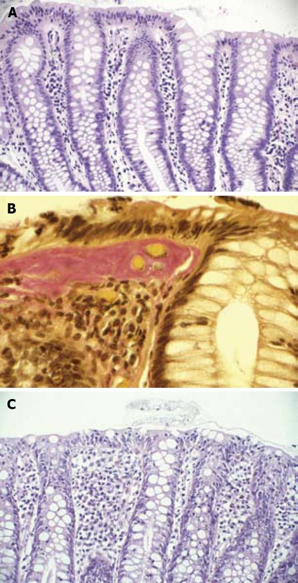
Figure 2 Biopsy from colon.
A: normal colonic mucosa (H&E stain); B: typical findings of CC-increased subepithelial collagen layer, inflammation of lamina propria and epithelial cell damage with intraepithelial lymphocytes (Van Gieson's stain); C: typical findings of LC-epithelial cell damage with intraepithelial lymphocytes and inflammation in the lamina propria (H&E stain).
- Citation: Tysk C, Bohr J, Nyhlin N, Wickbom A, Eriksson S. Diagnosis and management of microscopic colitis. World J Gastroenterol 2008; 14(48): 7280-7288
- URL: https://www.wjgnet.com/1007-9327/full/v14/i48/7280.htm
- DOI: https://dx.doi.org/10.3748/wjg.14.7280


