Copyright
©2008 The WJG Press and Baishideng.
World J Gastroenterol. Dec 21, 2008; 14(47): 7240-7246
Published online Dec 21, 2008. doi: 10.3748/wjg.14.7240
Published online Dec 21, 2008. doi: 10.3748/wjg.14.7240
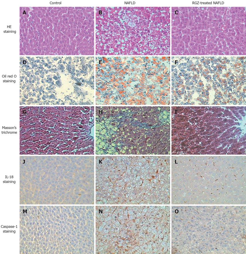
Figure 1 Histological studies of rat livers in normal control, NAFLD and RGZ-treated NAFLD groups (x 400).
Liver tissue sections were stained with HE, A: Normal control group; B: NAFLD group; C: RGZ-treated NAFLD group. Liver tissue sections were stained with oil red O, D: normal control group; E: NAFLD group; F: RGZ-treated NAFLD group. Liver tissue sections were stained with Masson’s trichrome, G: Normal control group; H: NAFLD group; I: RGZ-treated NAFLD group. Liver tissue sections were stained with immunohistochemistry for IL-18, J: Normal control group; K: NAFLD group; L: RGZ-treated NAFLD group. Liver tissue sections were stained with immunohistochemistry for caspase-1, M: Normal control group; N: NAFLD group; O: RGZ-treated NAFLD group. Histological changes in fatty liver disease and IL-18- or caspase-1-positive staining cells were rarely detectable in the control group. NAFLD rat liver showed steatosis and moderate inflammatory changes, fat droplet accumulation, mild fibrosis, strong IL-18- and caspase-1-positive staining. A significant improvement of steatosis, inflammation, fibrosis, and IL-18 and caspase-1 staining was observed in liver of the RGZ-treated NAFLD group.
- Citation: Wang HN, Wang YR, Liu GQ, Liu Z, Wu PX, Wei XL, Hong TP. Inhibition of hepatic interleukin-18 production by rosiglitazone in a rat model of nonalcoholic fatty liver disease. World J Gastroenterol 2008; 14(47): 7240-7246
- URL: https://www.wjgnet.com/1007-9327/full/v14/i47/7240.htm
- DOI: https://dx.doi.org/10.3748/wjg.14.7240


