Copyright
©2008 The WJG Press and Baishideng.
World J Gastroenterol. Dec 14, 2008; 14(46): 7093-7100
Published online Dec 14, 2008. doi: 10.3748/wjg.14.7093
Published online Dec 14, 2008. doi: 10.3748/wjg.14.7093
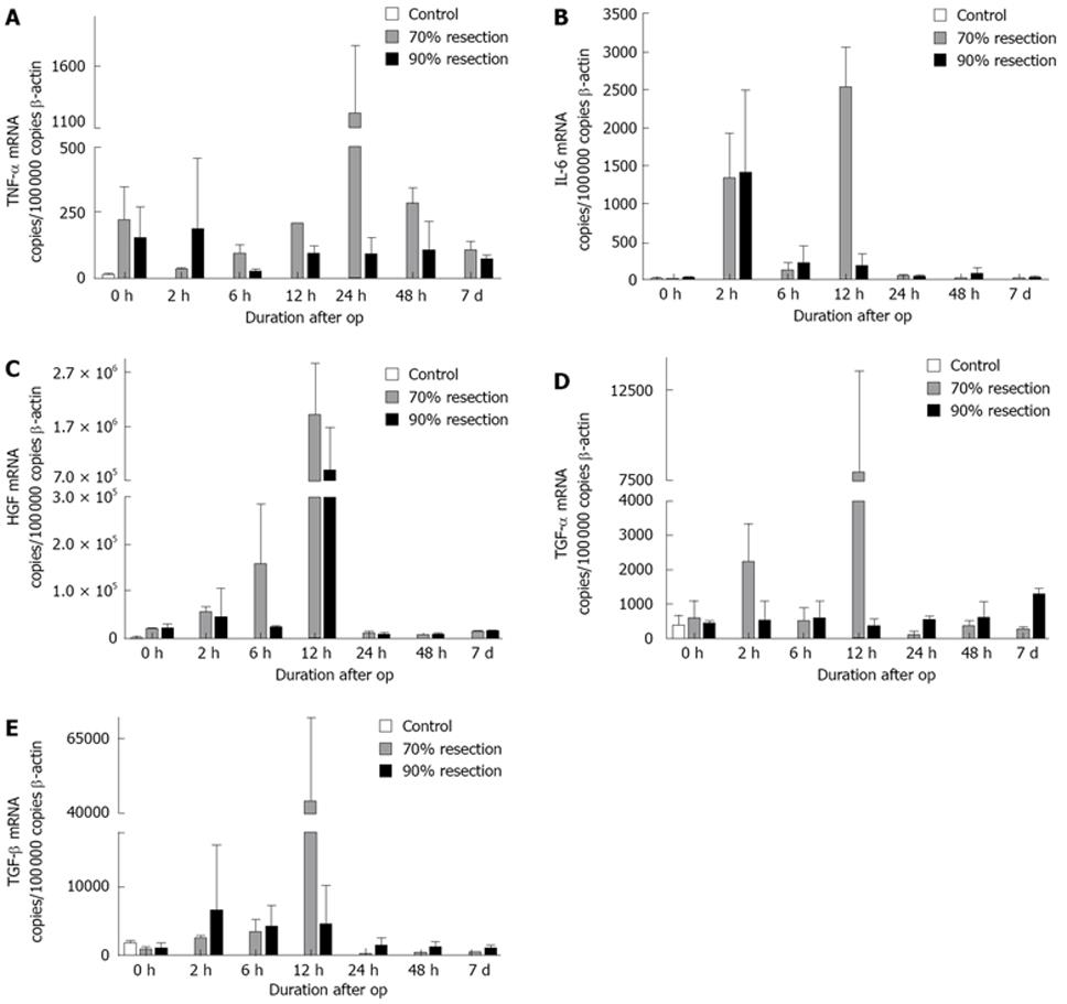
Figure 4 mRNA was isolated from regenerating liver tissue of rats at different time points after 70% or 90% resection, respectively.
A: TNF-α (< 50 copies/100 000 copies β-actin); B: IL-6 (< 15 copies/100 000 copies β-actin); C: HGF (50 000 copies/100 000 copies β-actin); D: TGF-β (< 2500 copies/100 000 copies β-actin); E: TGF-β (< 5500 copies/100 000 copies β-actin). Measurement of cytokine and growth-factor expression was performed by quantitative real-time (rt) PCR. Copy numbers of each gene were calculated from ct values. Data shown are the mean of four separate experiments with standard error of mean. All statistical significances were calculated against control animals. Baseline expression of each gene is given in parentheses.
- Citation: Sowa JP, Best J, Benko T, Bockhorn M, Gu Y, Niehues EM, Bucchi A, Benedetto-Castro EM, Gerken G, Rauen U, Schlaak JF. Extent of liver resection modulates the activation of transcription factors and the production of cytokines involved in liver regeneration. World J Gastroenterol 2008; 14(46): 7093-7100
- URL: https://www.wjgnet.com/1007-9327/full/v14/i46/7093.htm
- DOI: https://dx.doi.org/10.3748/wjg.14.7093


