Copyright
©2008 The WJG Press and Baishideng.
World J Gastroenterol. Dec 14, 2008; 14(46): 7033-7058
Published online Dec 14, 2008. doi: 10.3748/wjg.14.7033
Published online Dec 14, 2008. doi: 10.3748/wjg.14.7033
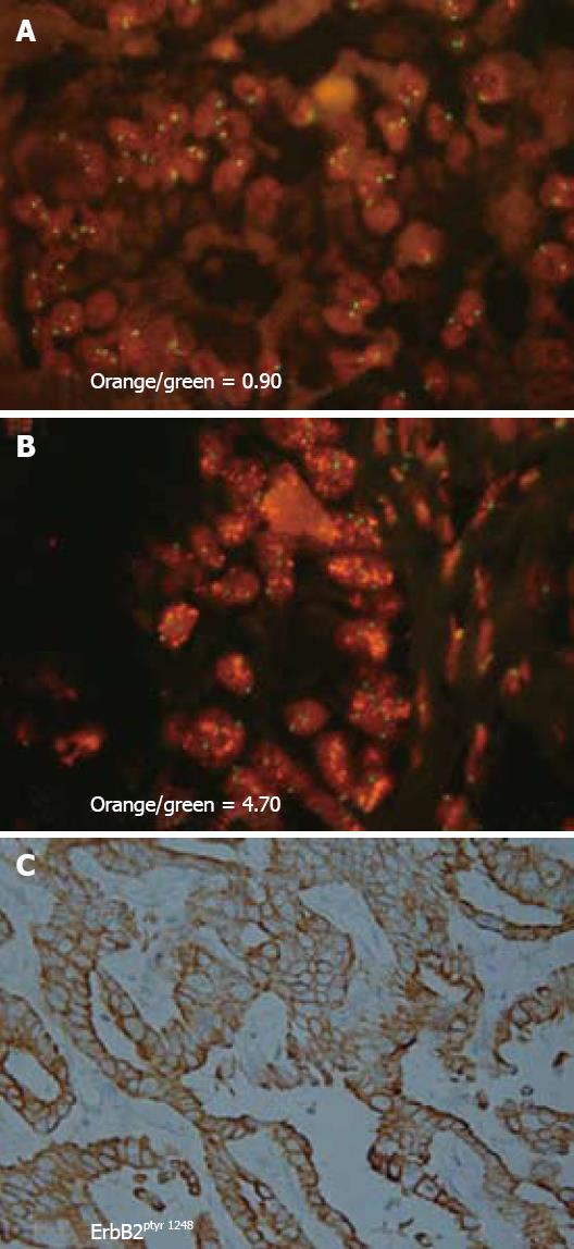
Figure 3 Representative photomicrographs demonstrating c-erbB2 gene amplification and a corresponding strong positive immunoreactivity for activated ErbB2 oncoprotein in the neoplastic cholangiocytes of a human intrahepatic cholangiocarcinoma.
A and B: Sections of two different intrahepatic cholangiocarcinomas with neoplastic cholangiocytes exhibiting FISH nuclear signals for c-erbB2 (orange dots) and for CEP17 (centromeric region of chromosome 17; green dots). Orange/green values indicate c-erbB2/CEP17 signal ratio for each cholangiocarcinoma. An orange/green ratio of > 2.0 = a tumor with c-erbB2 amplification (B), whereas a ratio value at or approximating 1.0 = a tumor without c-erbB2 amplification (A). The neoplastic cholangiocytes in the intrahepatic cholangiocarcinomas depicted in A (immunohistochemical staining not shown) and in B (with corresponding immunohistochemical staining shown in C) were each found to exhibit strong positive immunoreactivity at the plasma membrane for activated ErbB2 oncoprotein, using an antibody directed against phosphotyrosine 1248 of ErbB2. Tyrosine 1248 is the main autophosphorylation site in the carboxy terminal domain of ErbB2 linked to downstream signaling through the ras-raf-MEK-p42/44 MAPK signal transduction pathway. Thus, activation of ErbB2 via autophosphorylation at tyrosine 1248 in neoplastic cholangiocytes of intrahepatic cholangiocarcinomas can be demonstrated either in the presence or absence of c-erbB-2 amplification. (A, B × 330, C × 132).
- Citation: Sirica AE. Role of ErbB family receptor tyrosine kinases in intrahepatic cholangiocarcinoma. World J Gastroenterol 2008; 14(46): 7033-7058
- URL: https://www.wjgnet.com/1007-9327/full/v14/i46/7033.htm
- DOI: https://dx.doi.org/10.3748/wjg.14.7033


