Copyright
©2008 The WJG Press and Baishideng.
World J Gastroenterol. Nov 28, 2008; 14(44): 6863-6866
Published online Nov 28, 2008. doi: 10.3748/wjg.14.6863
Published online Nov 28, 2008. doi: 10.3748/wjg.14.6863
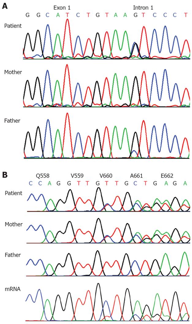
Figure 3 Diagram of the UBR1 gene mutations in our patient and her parents.
A: Exon 1-intron 1 transition showing a heterozygous nucleotide exchange at position +5 in the patient, IVS1+5G>C (c.81+G>C). Note the small peak in the father, indicating that he is mosaic for this mutation. B: Section of exon 17 showing a heterozygous 3 bp deletion in the patient and her mother, c.1978_1981delTTG (p.V660del), predicting the deletion of a highly conserved valine. In mRNA from lymphoblastoid cells from the patient the deletion is the predominant allele, indicating that mRNA from the allele with the splice site mutation is largely degradated.
-
Citation: Alkhouri N, Kaplan B, Kay M, Shealy A, Crowe C, Bauhuber S, Zenker M. Johanson-Blizzard syndrome with mild phenotypic features confirmed by
UBR1 gene testing. World J Gastroenterol 2008; 14(44): 6863-6866 - URL: https://www.wjgnet.com/1007-9327/full/v14/i44/6863.htm
- DOI: https://dx.doi.org/10.3748/wjg.14.6863


