Copyright
©2008 The WJG Press and Baishideng.
World J Gastroenterol. Nov 21, 2008; 14(43): 6743-6747
Published online Nov 21, 2008. doi: 10.3748/wjg.14.6743
Published online Nov 21, 2008. doi: 10.3748/wjg.14.6743
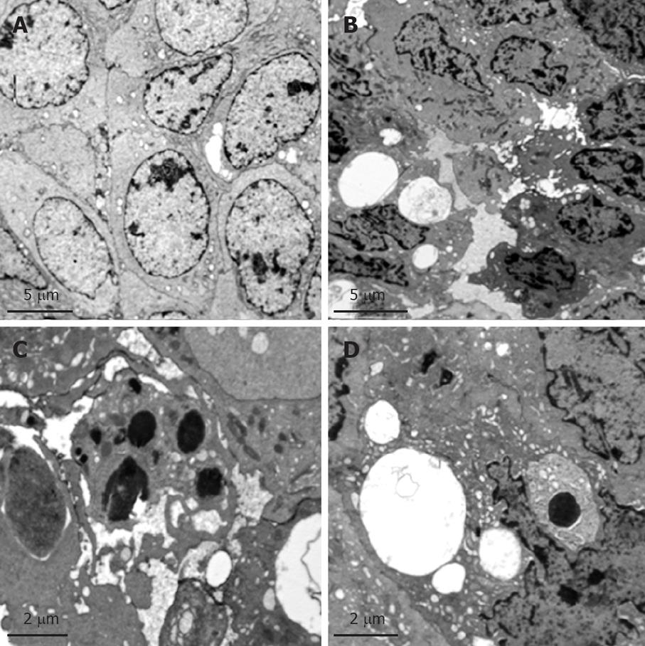
Figure 3 Ultrastructure of the targeted VX2 tumor tissues in sham group and PHIFU + UCA group under transmission electron microscope.
A: Tumor cells in sham group were pleomorphic or spindle-shaped of various sizes and with mitosis; nuclei were large and deformed, with clear nuclear membranes and rich euchromatin; chromatin particles were large with intranuclear pseudo-inclusions; there were multiple visible nucleoli (sham group, lead dyeing, × 3500); B: Tumor cells reduced in size; in some tumor cells, karyopyknosis was revealed, with chromatin margination and intercellular space widening (PHIFU + UCA group, lead dyeing, × 4000); C: High electron-density apoptotic bodies were present (PHIFU + UCA group, lead dyeing, × 8000); D: Vacuoles were present in cytoplasm of tumor cells (PHIFU + UCA group, lead dyeing, × 8000).
- Citation: Zhou CW, Li FQ, Qin Y, Liu CM, Zheng XL, Wang ZB. Non-thermal ablation of rabbit liver VX2 tumor by pulsed high intensity focused ultrasound with ultrasound contrast agent: Pathological characteristics. World J Gastroenterol 2008; 14(43): 6743-6747
- URL: https://www.wjgnet.com/1007-9327/full/v14/i43/6743.htm
- DOI: https://dx.doi.org/10.3748/wjg.14.6743


