Copyright
©2008 The WJG Press and Baishideng.
World J Gastroenterol. Nov 21, 2008; 14(43): 6681-6688
Published online Nov 21, 2008. doi: 10.3748/wjg.14.6681
Published online Nov 21, 2008. doi: 10.3748/wjg.14.6681
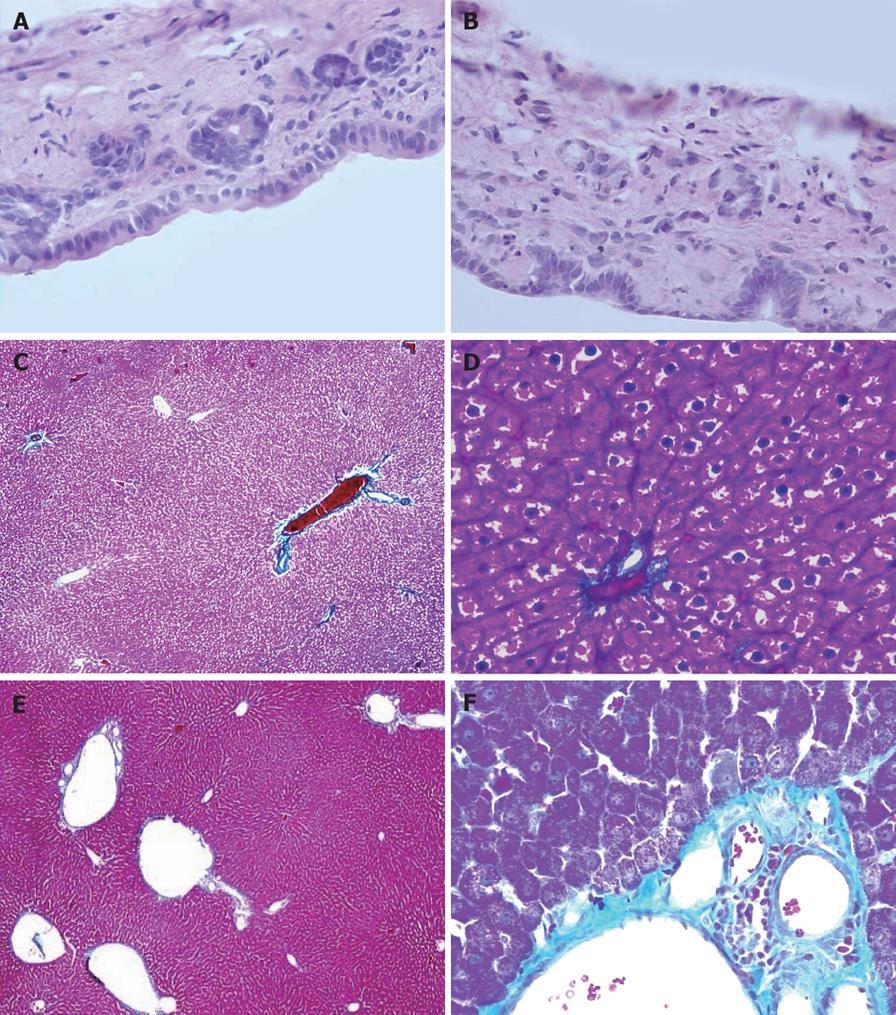
Figure 7 The pathological changes of hepatohilar bile duct and the liver in rats 6 mo after PVA using HE staining and Masson staining.
A: Hepatohilar bile ducts in control group (HE, × 400); B: Hepatohilar bile ducts in PVA group (HE, × 400). C: Masson staining of the liver 6 mo after operation in control group (× 40); D: Masson staining of the liver 6 mo after operation in control group (× 400). E: Masson staining of the liver 6 mo after PVA in PVA group (× 40); F: Masson staining of the liver 6 mo after PVA in PVA group (× 400).
- Citation: Li WG, Chen YL, Chen JX, Qu L, Xue BD, Peng ZH, Huang ZQ. Portal venous arterialization resulting in increased portal inflow and portal vein wall thickness in rats. World J Gastroenterol 2008; 14(43): 6681-6688
- URL: https://www.wjgnet.com/1007-9327/full/v14/i43/6681.htm
- DOI: https://dx.doi.org/10.3748/wjg.14.6681


