Copyright
©2008 The WJG Press and Baishideng.
World J Gastroenterol. Nov 14, 2008; 14(42): 6589-6592
Published online Nov 14, 2008. doi: 10.3748/wjg.14.6589
Published online Nov 14, 2008. doi: 10.3748/wjg.14.6589
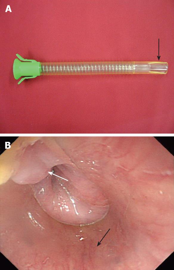
Figure 2 A fitted overtube.
A: A photograph of a fitted overtube. The distal end of a standard overtube is snipped off with a scissor in a rectangular form according to the size of the diverticular opening (arrow); B: An endoscopic view following insertion of a fitted overtube (white arrow: Tissue bridge; Black arrow: Overtube). The tissue bridge of the diverticulum is exposed in the fixed operational field. The surrounding esophageal wall is completely protected and stabilized.
- Citation: Lee CK, Chung IK, Park JY, Lee TH, Lee SH, Park SH, Kim HS, Kim SJ. Endoscopic diverticulotomy with an isolated-tip needle-knife papillotome (Iso-Tome) and a fitted overtube for the treatment of a Killian-Jamieson diverticulum. World J Gastroenterol 2008; 14(42): 6589-6592
- URL: https://www.wjgnet.com/1007-9327/full/v14/i42/6589.htm
- DOI: https://dx.doi.org/10.3748/wjg.14.6589


