Copyright
©2008 The WJG Press and Baishideng.
World J Gastroenterol. Nov 14, 2008; 14(42): 6589-6592
Published online Nov 14, 2008. doi: 10.3748/wjg.14.6589
Published online Nov 14, 2008. doi: 10.3748/wjg.14.6589
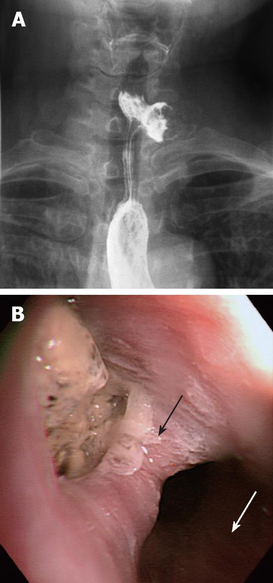
Figure 1 Barium swallow and endoscopic images.
A: An antero-posterior view of a barium swallow image, showing a diverticular sac filled with contrast and food debris in the left lateral side of the cervical esophagus; B: An endoscopic view of the KJD that originates on the anterolateral wall of the cervical esophagus, which was filled with impacted food debris (black arrow: the tissue bridge; white arrow: The esophageal lumen).
- Citation: Lee CK, Chung IK, Park JY, Lee TH, Lee SH, Park SH, Kim HS, Kim SJ. Endoscopic diverticulotomy with an isolated-tip needle-knife papillotome (Iso-Tome) and a fitted overtube for the treatment of a Killian-Jamieson diverticulum. World J Gastroenterol 2008; 14(42): 6589-6592
- URL: https://www.wjgnet.com/1007-9327/full/v14/i42/6589.htm
- DOI: https://dx.doi.org/10.3748/wjg.14.6589


