Copyright
©2008 The WJG Press and Baishideng.
World J Gastroenterol. Nov 14, 2008; 14(42): 6584-6588
Published online Nov 14, 2008. doi: 10.3748/wjg.14.6584
Published online Nov 14, 2008. doi: 10.3748/wjg.14.6584
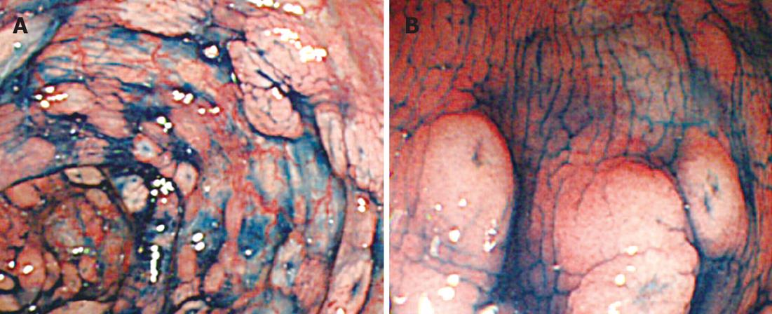
Figure 2 Colonoscopic images with indigo carmine dye.
A: Multiple lymphomatous polyposis was clearly depicted; B: Closer observation revealed polypoid and aphthoid lesions had tiny central depression.
- Citation: Hokama A, Tomoyose T, Yamamoto YI, Watanabe T, Hirata T, Kinjo F, Kato S, Ohshima K, Uezato H, Takasu N, Fujita J. Adult T-cell leukemia/lymphoma presenting multiple lymphomatous polyposis. World J Gastroenterol 2008; 14(42): 6584-6588
- URL: https://www.wjgnet.com/1007-9327/full/v14/i42/6584.htm
- DOI: https://dx.doi.org/10.3748/wjg.14.6584


