Copyright
©2008 The WJG Press and Baishideng.
World J Gastroenterol. Nov 7, 2008; 14(41): 6421-6424
Published online Nov 7, 2008. doi: 10.3748/wjg.14.6421
Published online Nov 7, 2008. doi: 10.3748/wjg.14.6421
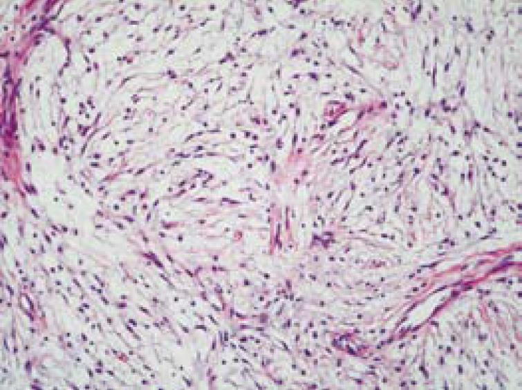
Figure 5 Histologic findings (hematoxylin and eosin stain, original magnification × 200).
The sections of the specimen are composed of fibrosis, focal sclerosis, plump spindle cells and vascular proliferation.
- Citation: Hsu CW, Lin CH, Yang TL, Chang HT. Splenic inflammatory pseudotumor mimicking angiosarcoma. World J Gastroenterol 2008; 14(41): 6421-6424
- URL: https://www.wjgnet.com/1007-9327/full/v14/i41/6421.htm
- DOI: https://dx.doi.org/10.3748/wjg.14.6421


