Copyright
©2008 The WJG Press and Baishideng.
World J Gastroenterol. Nov 7, 2008; 14(41): 6421-6424
Published online Nov 7, 2008. doi: 10.3748/wjg.14.6421
Published online Nov 7, 2008. doi: 10.3748/wjg.14.6421
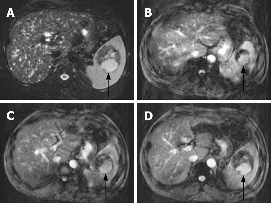
Figure 3 MRI.
A: In T2-weighted image, a well-circumscribed tumor (arrow) in the spleen, which showed partially dense intensity; B-D: In T1 contrast-enhancement dynamic study, peripheral nodule enhancement with gadolinium-contrast filling and pooling were noted in the tumor.
- Citation: Hsu CW, Lin CH, Yang TL, Chang HT. Splenic inflammatory pseudotumor mimicking angiosarcoma. World J Gastroenterol 2008; 14(41): 6421-6424
- URL: https://www.wjgnet.com/1007-9327/full/v14/i41/6421.htm
- DOI: https://dx.doi.org/10.3748/wjg.14.6421


