Copyright
©2008 The WJG Press and Baishideng.
World J Gastroenterol. Oct 28, 2008; 14(40): 6261-6264
Published online Oct 28, 2008. doi: 10.3748/wjg.14.6261
Published online Oct 28, 2008. doi: 10.3748/wjg.14.6261
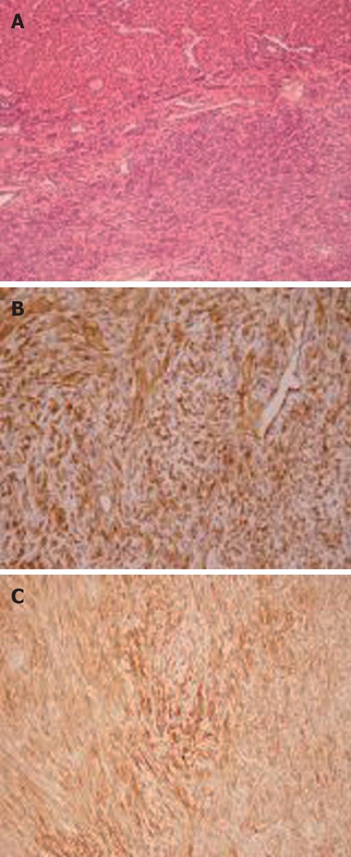
Figure 3 Tumor cells.
A: Microscopy showing a tumor composed of uniform collagen-forming spindle cells arranged in interlacing fascicles and well-encapsulated and differentiated from the adjacent non-cirrhotic liver parenchyma (HE, × 100); B: CD34 immunohistochemical staining demonstrating diffusely strong reactivity (× 200); C: Tumor cells showing diffuse immunohistochemical positivity for vimentin (× 200).
- Citation: Korkolis DP, Apostolaki K, Aggeli C, Plataniotis G, Gontikakis E, Volanaki D, Sebastiadou M, Dimitroulopoulos D, Xinopoulos D, Zografos GN, Vassilopoulos PP. Solitary fibrous tumor of the liver expressing CD34 and vimentin: A case report. World J Gastroenterol 2008; 14(40): 6261-6264
- URL: https://www.wjgnet.com/1007-9327/full/v14/i40/6261.htm
- DOI: https://dx.doi.org/10.3748/wjg.14.6261


