Copyright
©2008 The WJG Press and Baishideng.
World J Gastroenterol. Jan 28, 2008; 14(4): 607-611
Published online Jan 28, 2008. doi: 10.3748/wjg.14.607
Published online Jan 28, 2008. doi: 10.3748/wjg.14.607
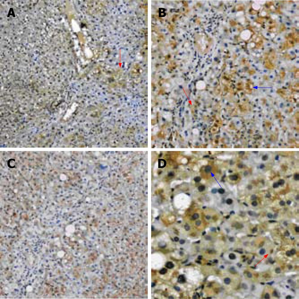
Figure 1 A: The liver tissue of mild CHB show the positive staining mainly distributed near the portal area (red arrow); B: The liver tissue of severe CHB show the positive staining is diffusely distributed; C: The positive staining mainly involves the hepatocytes (blue arrow), only a few of liver interstitial cells are involved (red arrow); D: The positive staining is mainly distributed in the plasma (blue arrow), some on the membrane.
occasionally in the inclusion bodies (red arrow). (A, B: Immunohistochemistry SABC × 100; C: Immunohistochemistry SABC × 200; D: Immunohistochemistry SABC × 400).
- Citation: Zhao ZX, Cai QX, Peng XM, Chong YT, Gao ZL. Expression of SOCS-1 in the liver tissues of chronic hepatitis B and its clinical significance. World J Gastroenterol 2008; 14(4): 607-611
- URL: https://www.wjgnet.com/1007-9327/full/v14/i4/607.htm
- DOI: https://dx.doi.org/10.3748/wjg.14.607


