Copyright
©2008 The WJG Press and Baishideng.
World J Gastroenterol. Oct 21, 2008; 14(39): 6036-6043
Published online Oct 21, 2008. doi: 10.3748/wjg.14.6036
Published online Oct 21, 2008. doi: 10.3748/wjg.14.6036
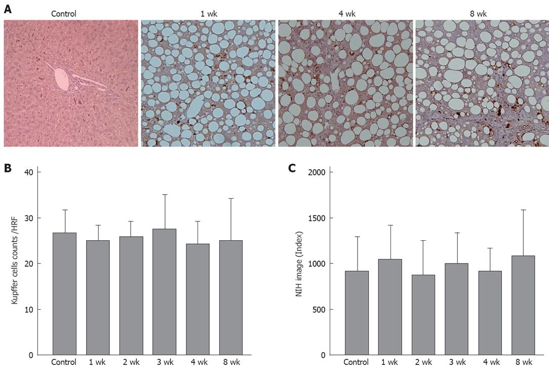
Figure 6 A: Kupffer cell immunohistochemical staining (× 200).
Brown-stained cells are positive; B: There were no significant differences between the different groups in the number of stained cell per field; C: No significant differences were found between the groups in the quantitative analyses (n = 5).
- Citation: Tsujimoto T, Kawaratani H, Kitazawa T, Hirai T, Ohishi H, Kitade M, Yoshiji H, Uemura M, Fukui H. Decreased phagocytic activity of Kupffer cells in a rat nonalcoholic steatohepatitis model. World J Gastroenterol 2008; 14(39): 6036-6043
- URL: https://www.wjgnet.com/1007-9327/full/v14/i39/6036.htm
- DOI: https://dx.doi.org/10.3748/wjg.14.6036


