Copyright
©2008 The WJG Press and Baishideng.
World J Gastroenterol. Oct 21, 2008; 14(39): 6036-6043
Published online Oct 21, 2008. doi: 10.3748/wjg.14.6036
Published online Oct 21, 2008. doi: 10.3748/wjg.14.6036
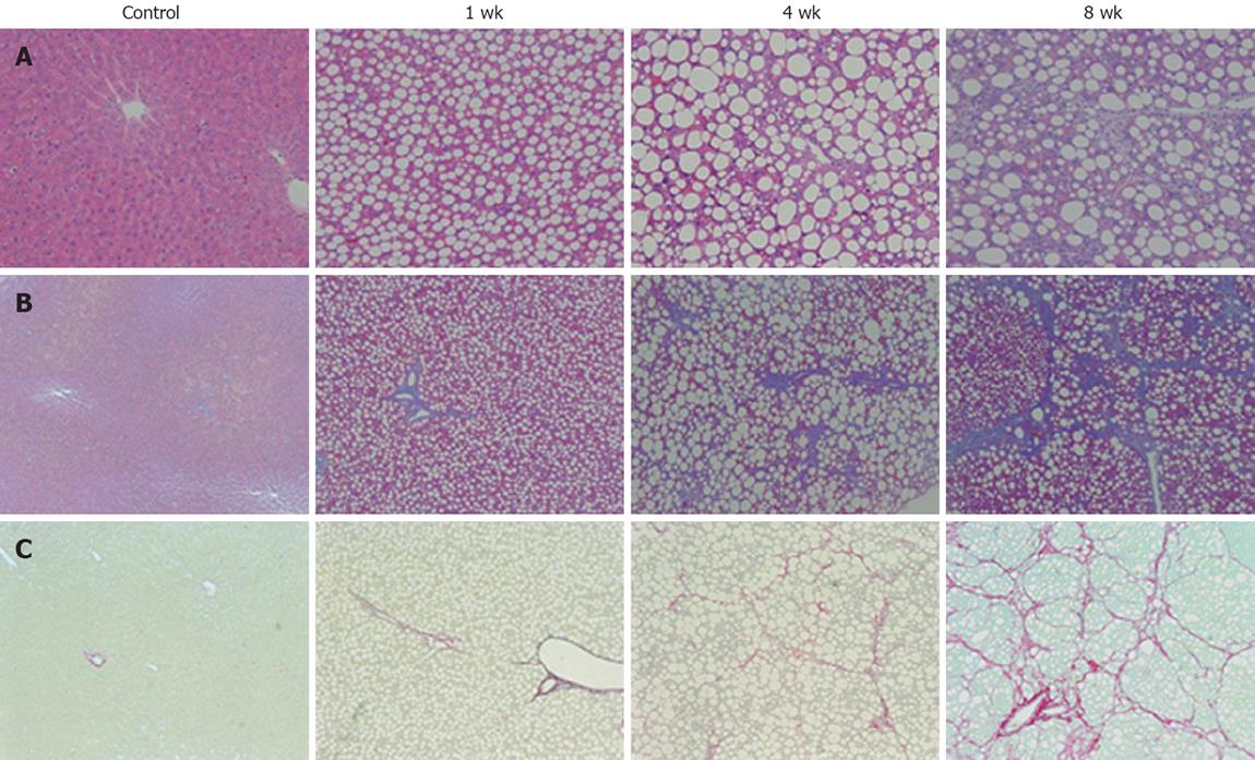
Figure 2 Histological analysis of the liver sections.
A: Hematoxylin and eosin stain (× 200); B: Azan stain (× 100); C: Sirius red stain (× 100). Histological examination of the liver tissue of 1-wk CDAA-fed rats showed inflammation and fat deposits, but no fibrosis, corresponding to Matteoni’s type 2. The 4-wk CDAA-fed rats had more inflammation, fat deposits, and fibrosis, which was equivalent to Matteoni’s type 3 and grade 2/stage 2 of Brunt’s NASH classification. The histological findings in the 8-wk CDAA-fed rats were equivalent to Matteoni’s type 4 and Brunt’s grade 2/stage 3. In the 4 and 8-wk groups, Sirius red staining revealed abundant collagen (n = 5).
- Citation: Tsujimoto T, Kawaratani H, Kitazawa T, Hirai T, Ohishi H, Kitade M, Yoshiji H, Uemura M, Fukui H. Decreased phagocytic activity of Kupffer cells in a rat nonalcoholic steatohepatitis model. World J Gastroenterol 2008; 14(39): 6036-6043
- URL: https://www.wjgnet.com/1007-9327/full/v14/i39/6036.htm
- DOI: https://dx.doi.org/10.3748/wjg.14.6036


