Copyright
©2008 The WJG Press and Baishideng.
World J Gastroenterol. Oct 21, 2008; 14(39): 5980-5989
Published online Oct 21, 2008. doi: 10.3748/wjg.14.5980
Published online Oct 21, 2008. doi: 10.3748/wjg.14.5980
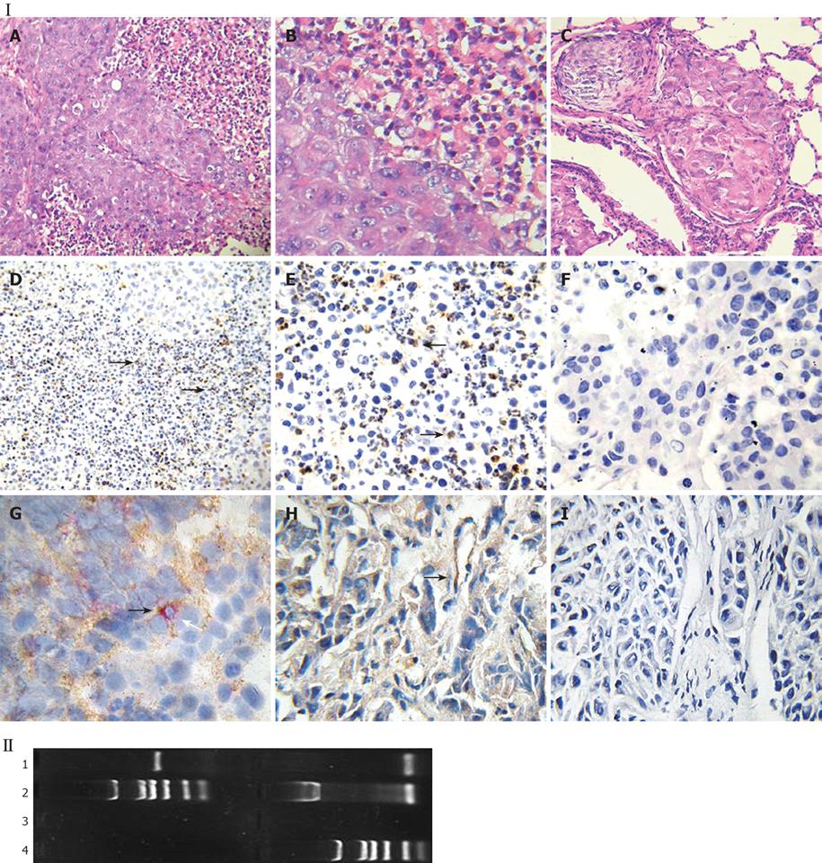
Figure 4 Fgl2 expression evidenced in mouse tumor tissue.
Male BALB/c-nu/nu mice were subcutaneously injected MHCC97LM6 cell lines and tumor tissues were harvested 36 d later. Panel I: A-C, HE staining in tumor tissue at injection site (A, × 200; B, × 400) and lung metastatic tumor tissue (C, × 200); D-I: mfgl2 expression in tumor tissue of human hepatocellular carcinoma (HCC) nude mice model (D, SP × 200; E, SP × 400; G, dual staining of mfgl2 and marker of macrophages; H, dual staining of mfgl2 and marker of endothelial cells; F and I, negative controls). Arrows in D and E indicate fgl2 positive cells, arrows in G and F indicate the fgl2 positive macrophage and endothelial cells, respectively. Panel II: mfgl2 mRNA expression in tumor tissue (1), PCDNA3.1-fgl2 plasmid as positive control (2), PCDNA3.1 as negative control (3) and DL-2000 marker (4).
-
Citation: Su K, Chen F, Yan WM, Zeng QL, Xu L, Xi D, Pi B, Luo XP, Ning Q. Fibrinogen-like protein 2/fibroleukin prothrombinase contributes to tumor hypercoagulability
via IL-2 and IFN-γ. World J Gastroenterol 2008; 14(39): 5980-5989 - URL: https://www.wjgnet.com/1007-9327/full/v14/i39/5980.htm
- DOI: https://dx.doi.org/10.3748/wjg.14.5980


