Copyright
©2008 The WJG Press and Baishideng.
World J Gastroenterol. Oct 14, 2008; 14(38): 5834-5841
Published online Oct 14, 2008. doi: 10.3748/wjg.14.5834
Published online Oct 14, 2008. doi: 10.3748/wjg.14.5834
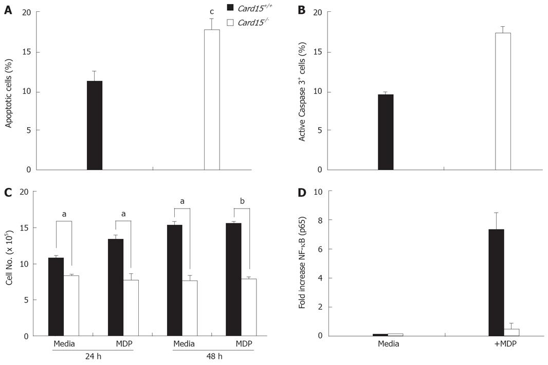
Figure 4 Decreased survival of Nod2-/- CECs in vitro.
A: The level of apoptotic cells in cultures of primary CECs from Nod2+/+ and Nod2-/- mice was determined by annexin V and propidium iodide (PI) staining and flow cytometry. Data was collected from 5 experiments and shows the mean (± SE) values. cP < 0.01 B: The frequency of Nod2+/+ and Nod2-/- CECs containing caspase3 activity was determined using the flow cytometry based NucViewTM488 Caspase-3 assay. The data represents the mean (± SE) values collected from two experiments. C: The growth of Nod2+/+ and Nod2-/- CECs in the absence (Media) or presence of 10 μg/mL muramyl dipeptide (MDP) was determined by comparing the number of viable CEC at the initiation of culture and after 24 h and 48 h. The data shown represents the mean (± SE) of 3 experiments. aP < 0.05, bP < 0.001. D: NF-κB activation in Nod2+/+ (filled bars) and Nod2-/- (open bars) CEC after exposure to muramyl dipeptide (MDP) was determined by quantitating NF-κB p65 levels in nuclear extracts using a Transfactor Kit as described in Methods section. Values were normalized to control values of cells grown in media alone. The data shown represents the mean (± SE) values from 3 independent experiments.
- Citation: Cruickshank SM, Wakenshaw L, Cardone J, Howdle PD, Murray PJ, Carding SR. Evidence for the involvement of NOD2 in regulating colonic epithelial cell growth and survival. World J Gastroenterol 2008; 14(38): 5834-5841
- URL: https://www.wjgnet.com/1007-9327/full/v14/i38/5834.htm
- DOI: https://dx.doi.org/10.3748/wjg.14.5834


