Copyright
©2008 The WJG Press and Baishideng.
World J Gastroenterol. Oct 14, 2008; 14(38): 5834-5841
Published online Oct 14, 2008. doi: 10.3748/wjg.14.5834
Published online Oct 14, 2008. doi: 10.3748/wjg.14.5834
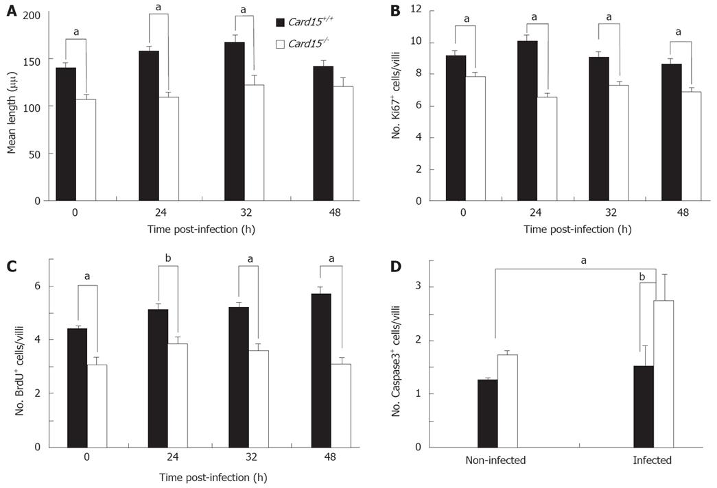
Figure 3 Decreased proliferation of Nod2-/- CECs in vivo.
A: Mean crypt-villous height measurements were determined by measuring distances from the base to the tip of the crypt of at least 20 crypts from sections taken at the distal, mid and proximal parts of the colon of Nod2+/+ and Nod2-/- mice (n = 5) prior to (0 h) and at different times after peroral Salmonella infection. The mean (± SE) of counting at least 20 crypts in 3-4 sections of 4-5 mice of each strain is shown. B: Sections of Nod2+/+ and Nod2-/- colon were stained with anti-Ki67 antibody with the mean (± SE) number of stained cells in 5-6 sections from three mice of each strain shown. aP < 0.05. C: BrdU-incorporation was assessed by injecting Nod2+/+ and Nod2-/- mice with BrdU 1 h prior to removing the colons, which were then sectioned and stained with an anti-BrdU antibody. The graph represents the mean (± SE) number of BrdU+ epithelial cells per crypt as determined by counting stained cells in > 20 crypts from 5-6 sections taken from proximal, mid and distal regions of the colons from three mice of each strain. aP < 0.05, bP < 0.001. D: Sections of colon from Nod2+/+ and Nod2-/- mice were stained with anti-active caspase3 antibodies with the mean (± SE) number of caspase3+ cells in 4-5 sections from three mice of each strain shown. aP < 0.05, bP < 0.001.
- Citation: Cruickshank SM, Wakenshaw L, Cardone J, Howdle PD, Murray PJ, Carding SR. Evidence for the involvement of NOD2 in regulating colonic epithelial cell growth and survival. World J Gastroenterol 2008; 14(38): 5834-5841
- URL: https://www.wjgnet.com/1007-9327/full/v14/i38/5834.htm
- DOI: https://dx.doi.org/10.3748/wjg.14.5834


