Copyright
©2008 The WJG Press and Baishideng.
World J Gastroenterol. Oct 14, 2008; 14(38): 5797-5809
Published online Oct 14, 2008. doi: 10.3748/wjg.14.5797
Published online Oct 14, 2008. doi: 10.3748/wjg.14.5797
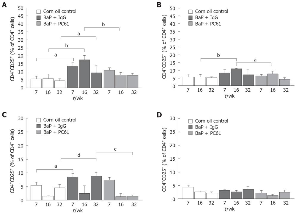
Figure 6 CD4+CD25+ T cells in distinct microenvironments.
Lymphocytes in RLNs (A), PLNs (B), spleen (C), and thymus (D) were double-stained with FITC-anti-CD25 and PE-anti-CD4 mAbs and quantified by flow cytometry in the control, BaP + IgG-treated, and BaP + PC61-treated mice at wk 7, wk 16, and wk 32. aP < 0.05, bP < 0.01, cP < 0.005, dP < 0.001 vs the control mice.
- Citation: Chen YL, Fang JH, Lai MD, Shan YS. Depletion of CD4+CD25+ regulatory T cells can promote local immunity to suppress tumor growth in benzo[a]pyrene-induced forestomach carcinoma. World J Gastroenterol 2008; 14(38): 5797-5809
- URL: https://www.wjgnet.com/1007-9327/full/v14/i38/5797.htm
- DOI: https://dx.doi.org/10.3748/wjg.14.5797


