Copyright
©2008 The WJG Press and Baishideng.
World J Gastroenterol. Oct 7, 2008; 14(37): 5755-5759
Published online Oct 7, 2008. doi: 10.3748/wjg.14.5755
Published online Oct 7, 2008. doi: 10.3748/wjg.14.5755
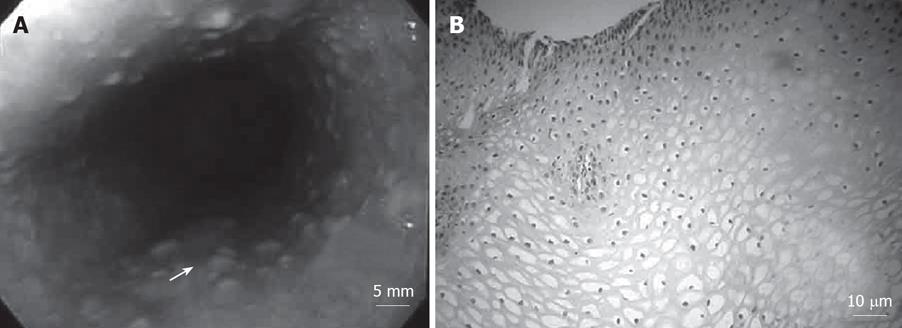
Figure 4 Polypoid lesions in the esophagus.
A: Endoscopy of the upper digestive tract showing whitish polypoid lesions in the esophagus and macroscopic appearance of smooth lesions. The size of all these polyps was within 5 mm; B: Pathological appearance of the esophagus. Histlogically, a specimen confirmed the diagnosis of glycogenic acanthosis.
- Citation: Umemura K, Takagi S, Ishigaki Y, Iwabuchi M, Kuroki S, Kinouchi Y, Shimosegawa T. Gastrointestinal polyposis with esophageal polyposis is useful for early diagnosis of Cowden’s disease. World J Gastroenterol 2008; 14(37): 5755-5759
- URL: https://www.wjgnet.com/1007-9327/full/v14/i37/5755.htm
- DOI: https://dx.doi.org/10.3748/wjg.14.5755


