Copyright
©2008 The WJG Press and Baishideng.
World J Gastroenterol. Oct 7, 2008; 14(37): 5749-5754
Published online Oct 7, 2008. doi: 10.3748/wjg.14.5749
Published online Oct 7, 2008. doi: 10.3748/wjg.14.5749
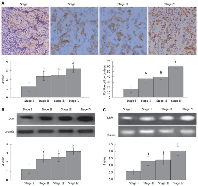
Figure 1 A: The expression of Livin was measured by IHC (SP × 400).
Optical density value and positive cell percentage in clinicopathologic stage two, three and four was significantly higher than that of stage one (bP < 0.01). Furthermore, optical density value and positive cell percentage in clinicopathologic stage four was significantly higher than that of stage two and three (dP < 0.01). IHC showed that Livin had significant expression in the cytoplasm and nucleus in the Stage II, III and IV (the cytoplasm and nucleus had stained yellow), slight expression in the cytoplasm and nucleus in Stage I (the cytoplasm and nucleus had slightly stained yellow); B: The expression of Livin by Western blotting showed that expression of Livin in clinicopathologic stage two, three and four was significantly higher than that of stage one (fP < 0.01). Optical density value and positive cell percentage in clinicopathologic stage four was significantly higher than that of stage two and three (hP < 0.01); C: mRNA level of Livin was tested by RT-PCR. Up-regulation of Livin gene transcription matched with the protein level of Livin that was significantly increased along with the progression of esophageal carcinoma. Optical density value in clinicopathologic stage two, three and four was significantly higher than that of stage one (jP < 0.01). Optical density value and positive cell percentage in clinicopathologic stage four was significantly higher than that of stage two and three (lP < 0.01).
- Citation: Chen L, Ren GS, Li F, Sun SQ. Expression of Livin and vascular endothelial growth factor in different clinical stages of human esophageal carcinoma. World J Gastroenterol 2008; 14(37): 5749-5754
- URL: https://www.wjgnet.com/1007-9327/full/v14/i37/5749.htm
- DOI: https://dx.doi.org/10.3748/wjg.14.5749


