Copyright
©2008 The WJG Press and Baishideng.
World J Gastroenterol. Sep 28, 2008; 14(36): 5564-5569
Published online Sep 28, 2008. doi: 10.3748/wjg.14.5564
Published online Sep 28, 2008. doi: 10.3748/wjg.14.5564
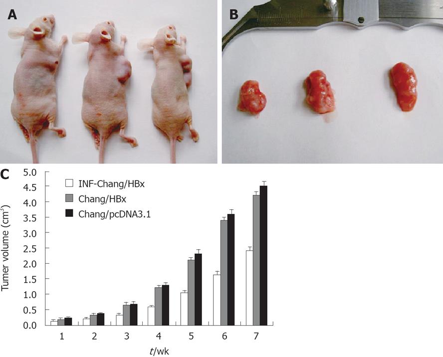
Figure 5 A: A nude mouse migration model with pcDNA3.
1-HBx Chang cells, IFN-α-Chang/HBx cells and Chang/pcDNA3.1 cells were constructed on the 7th wk; B: The volume of neoplasms in the three groups were observed. The sizes of the hepatomas from Chang/pcDNA3.1 and IFN-α-Chang/HBx injected nude mice were obviously larger than the tumors of Chang/HBx injected mice; C: Neoplasm growth in the Chang/HBx-inoculated nude mice compared between IFN-α-Chang/HBx and the control Chang/pcDNA3.1-inoculated mice.
- Citation: Yang JQ, Pan GD, Chu GP, Liu Z, Liu Q, Xiao Y, Yuan L. Interferon-alpha restrains growth and invasive potential of hepatocellular carcinoma induced by hepatitis B virus X protein. World J Gastroenterol 2008; 14(36): 5564-5569
- URL: https://www.wjgnet.com/1007-9327/full/v14/i36/5564.htm
- DOI: https://dx.doi.org/10.3748/wjg.14.5564


