Copyright
©2008 The WJG Press and Baishideng.
World J Gastroenterol. Sep 28, 2008; 14(36): 5549-5556
Published online Sep 28, 2008. doi: 10.3748/wjg.14.5549
Published online Sep 28, 2008. doi: 10.3748/wjg.14.5549
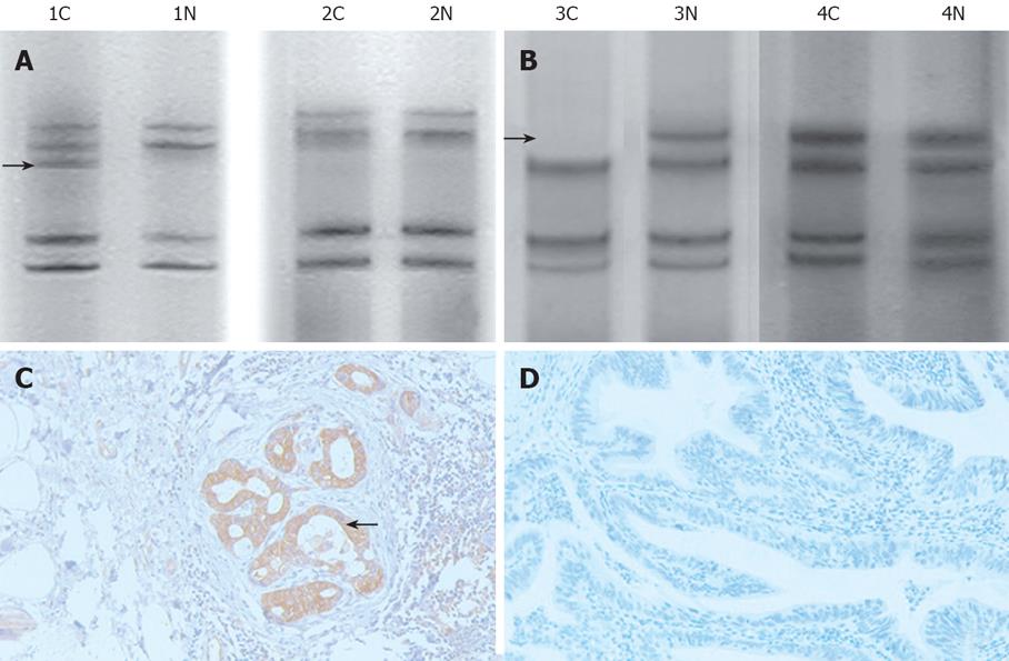
Figure 4 PAGE of D17S396 locus and immunohistochemistry of nm23H1 protein in gallbladder carcinoma.
A: No difference in tumor tissue (2C) and normal tissue (2N). MSI was positive when an allele band was added (arrow) in tumor tissue (1C) as compared with normal tissue (1N); B: No difference in tumor tissue (4C) and normal tissue (4N). LOH was positive in the absence of an allele band (arrow) in tumor tissue (3C) as compared with normal tissue (3N); C: Brown-yellow granules of nm23H1 protein mostly located in cytoplasm, and part of the stained nucleolus and membrane of cells were observed (arrow, × 200); D: Control group, PBS replaced anti-nm23H1 protein as the first antibody (× 200).
- Citation: Yang YQ, Wu L, Chen JX, Sun JZ, Li M, Li DM, Lu HY, Su ZH, Lin XQ, Li JC. Relationship between nm23H1 genetic instability and clinical pathological characteristics in Chinese digestive system cancer patients. World J Gastroenterol 2008; 14(36): 5549-5556
- URL: https://www.wjgnet.com/1007-9327/full/v14/i36/5549.htm
- DOI: https://dx.doi.org/10.3748/wjg.14.5549


