Copyright
©2008 The WJG Press and Baishideng.
World J Gastroenterol. Aug 21, 2008; 14(31): 4968-4971
Published online Aug 21, 2008. doi: 10.3748/wjg.14.4968
Published online Aug 21, 2008. doi: 10.3748/wjg.14.4968
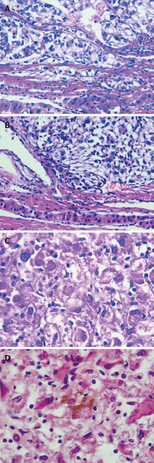
Figure 3 Microscopy showing diffusely infiltrating tumor cells (A) and fibrous tissues (B) between the lesion and normal hepatocytes with a few broken tumor cells through the capsule infiltrating the adjacent liver tissue (× 200), pleomorphic tumor cells with round, spindle-shaped and irregular morphologies(× 400) (C), and some tumor cells containing melanin deposition (× 200) (D).
- Citation: Gong L, Li YH, Zhao JY, Wang XX, Zhu SJ, Zhang W. Primary malignant melanoma of the liver: A case report. World J Gastroenterol 2008; 14(31): 4968-4971
- URL: https://www.wjgnet.com/1007-9327/full/v14/i31/4968.htm
- DOI: https://dx.doi.org/10.3748/wjg.14.4968


