Copyright
©2008 The WJG Press and Baishideng.
World J Gastroenterol. Aug 21, 2008; 14(31): 4867-4872
Published online Aug 21, 2008. doi: 10.3748/wjg.14.4867
Published online Aug 21, 2008. doi: 10.3748/wjg.14.4867
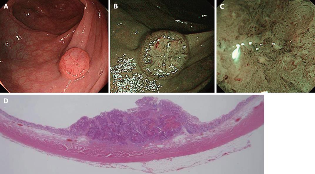
Figure 4 NBI image of colorectal cancer.
A: Conventional view of an IIa + IIc lesion, 12 mm in size, located in the transverse colon; B: NBI view shows a well demarcated area and meshed capillary vessels clearly visible characterized by thick diameter, branching and curtail irregularity; C: Magnifying NBI view additionally shows the presence of a nearly avascular or loose microvascular area due to histological desmoplastic changes in the stromal tissue, suggesting deep submucosal invasion; D: Histopathological analysis revealed an adenocarcinoma invading deeply into the submucosa (2500 μm) with lymphovascular invasion.
- Citation: Emura F, Saito Y, Ikematsu H. Narrow-band imaging optical chromocolonoscopy: Advantages and limitations. World J Gastroenterol 2008; 14(31): 4867-4872
- URL: https://www.wjgnet.com/1007-9327/full/v14/i31/4867.htm
- DOI: https://dx.doi.org/10.3748/wjg.14.4867


