Copyright
©2008 The WJG Press and Baishideng.
World J Gastroenterol. Aug 14, 2008; 14(30): 4763-4770
Published online Aug 14, 2008. doi: 10.3748/wjg.14.4763
Published online Aug 14, 2008. doi: 10.3748/wjg.14.4763
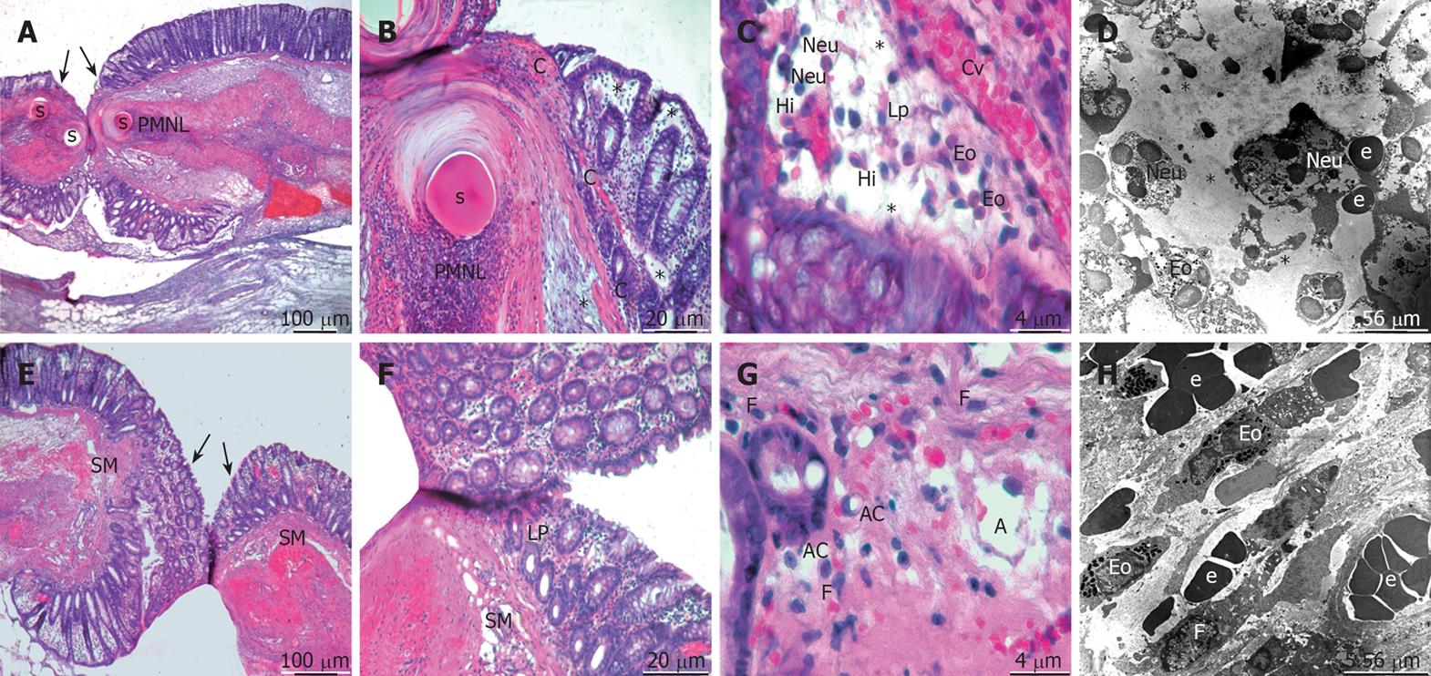
Figure 1 An overview of healing at day 1 after operation.
The micrographs “A-C, E-G” represent hematoxylin-eosin stained sections and “D, H” T.E.M. images of the control (upper panels) and propolis (bottom panels) group. Arrow: the anastomosis cite; Asterisk: Edema; PMNL: Polymorphonuclear leucocytes; s: Suture material; C: Congestion; Cv: Capillary vessel; LP: Lamina propria; Eo: Eosinophil; Neu: Neutrophil; Hi: Histiocyte; e: Eritrocyte; SM: Submucosa; AC: Apoptotic cell; F: Fibroblast; A: Angiogenesis.
- Citation: Kilicoglu SS, Kilicoglu B, Erdemli E. Ultrastructural view of colon anastomosis under propolis effect by transmission electron microscopy. World J Gastroenterol 2008; 14(30): 4763-4770
- URL: https://www.wjgnet.com/1007-9327/full/v14/i30/4763.htm
- DOI: https://dx.doi.org/10.3748/wjg.14.4763


