Copyright
©2008 The WJG Press and Baishideng.
World J Gastroenterol. Jan 21, 2008; 14(3): 448-453
Published online Jan 21, 2008. doi: 10.3748/wjg.14.448
Published online Jan 21, 2008. doi: 10.3748/wjg.14.448
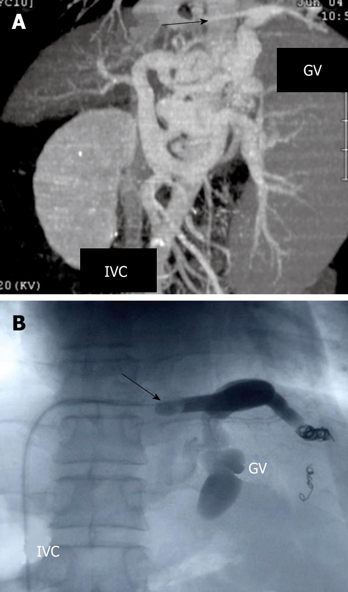
Figure 1 Multidimensional computed tomography (MDCT) revealing the varices supplied by the left gastric vein and drained into the subphrenic vein (arrow) which is connected to the inferior vena cava (IVC) (A), and fluoroscopic image of BRTO showing placement of the occlusive balloon catheter in the subphrenic vein (arrow) (B).
After the blood vessels from the GV except for the main drainage vein were blocked with coils, 5% solution of ethanolamine oleate with iopamidol (EOI) was injected into the varices.
- Citation: Kameda N, Higuchi K, Shiba M, Kadouchi K, Machida H, Okazaki H, Tanigawa T, Watanabe T, Tominaga K, Fujiwara Y, Nakamura K, Arakawa T. Management of gastric fundal varices without gastro-renal shunt in 15 patients. World J Gastroenterol 2008; 14(3): 448-453
- URL: https://www.wjgnet.com/1007-9327/full/v14/i3/448.htm
- DOI: https://dx.doi.org/10.3748/wjg.14.448


