Copyright
©2008 The WJG Press and Baishideng.
World J Gastroenterol. Aug 7, 2008; 14(29): 4709-4712
Published online Aug 7, 2008. doi: 10.3748/wjg.14.4709
Published online Aug 7, 2008. doi: 10.3748/wjg.14.4709
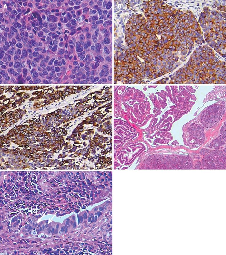
Figure 3 Histopathological findings.
A: The tumor cells are composed predominantly of small to medium-sized, round or oval cells with hyperchromatic nuclei, inconspicuous nucleoli and scanty cytoplasm (HE, × 400); Immunohistochemically, the tumor cells are positive for synaptophysin (B) and low-molecular-weight cytokeratin (C) (× 200); D: Light microscopy shows the coexistence of villous adenoma and small cell neuroendocrine carcinoma (HE, × 20); E: The epithelium of the adenoma (long arrows) is in continuity with the small cell neuroendocrine carcinoma (short arrows) (HE, × 200).
- Citation: Sun JH, Chao M, Zhang SZ, Zhang GQ, Li B, Wu JJ. Coexistence of small cell neuroendocrine carcinoma and villous adenoma in the ampulla of Vater. World J Gastroenterol 2008; 14(29): 4709-4712
- URL: https://www.wjgnet.com/1007-9327/full/v14/i29/4709.htm
- DOI: https://dx.doi.org/10.3748/wjg.14.4709


