Copyright
©2008 The WJG Press and Baishideng.
World J Gastroenterol. Aug 7, 2008; 14(29): 4709-4712
Published online Aug 7, 2008. doi: 10.3748/wjg.14.4709
Published online Aug 7, 2008. doi: 10.3748/wjg.14.4709
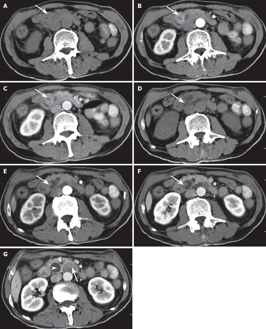
Figure 1 Abdominal CT reveals two well-defined masses in the ampulla of Vater.
On precontrast CT scanning, the two masses (arrow) show isoattenuation to the surrounding pancreatic parenchyma (A and D). On arterial and portal phase images after contrast enhancement, the larger oval mass (arrow) shows slightly higher attenuation than the surrounding pancreas (B and C). The smaller polypous mass (arrow) with a long pedicle and stalk shows slightly lower attenuation than the surrounding pancreas (E and F). Contrast-enhanced CT scan (G) shows peripancreatic lymphadenopathy (arrow) and dilatation of the common bile duct and the pancreatic duct (arrowheads).
- Citation: Sun JH, Chao M, Zhang SZ, Zhang GQ, Li B, Wu JJ. Coexistence of small cell neuroendocrine carcinoma and villous adenoma in the ampulla of Vater. World J Gastroenterol 2008; 14(29): 4709-4712
- URL: https://www.wjgnet.com/1007-9327/full/v14/i29/4709.htm
- DOI: https://dx.doi.org/10.3748/wjg.14.4709


