Copyright
©2008 The WJG Press and Baishideng.
World J Gastroenterol. Aug 7, 2008; 14(29): 4701-4704
Published online Aug 7, 2008. doi: 10.3748/wjg.14.4701
Published online Aug 7, 2008. doi: 10.3748/wjg.14.4701
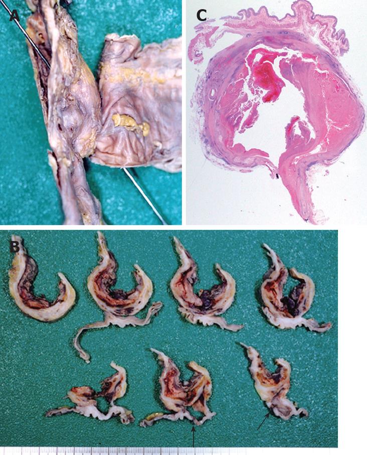
Figure 6 Formalin-fixed duodenum and aorta specimen demonstrating a sonde inserted from the rupture on the duodenal mucosa to the inside of the aortic lumen (A), horizontal cross sections showing a firm adhesion between the aorta and its wall as well as a rupture (black arrow) on the third part of the duodenum (B); and a microphotograph of a horizontal cross section showing an adhesion between the duodenum and the aorta with atherosclerosis-contained clotted blood (C).
The histology of the duodenal appears normal (HE, × 5).
- Citation: Ihama Y, Miyazaki T, Fuke C, Ihama Y, Matayoshi R, Kohatsu H, Kinjo F. An autopsy case of a primary aortoenteric fistula: A pitfall of the endoscopic diagnosis. World J Gastroenterol 2008; 14(29): 4701-4704
- URL: https://www.wjgnet.com/1007-9327/full/v14/i29/4701.htm
- DOI: https://dx.doi.org/10.3748/wjg.14.4701


