Copyright
©2008 The WJG Press and Baishidengs.
World J Gastroenterol. Aug 7, 2008; 14(29): 4627-4632
Published online Aug 7, 2008. doi: 10.3748/wjg.14.4627
Published online Aug 7, 2008. doi: 10.3748/wjg.14.4627
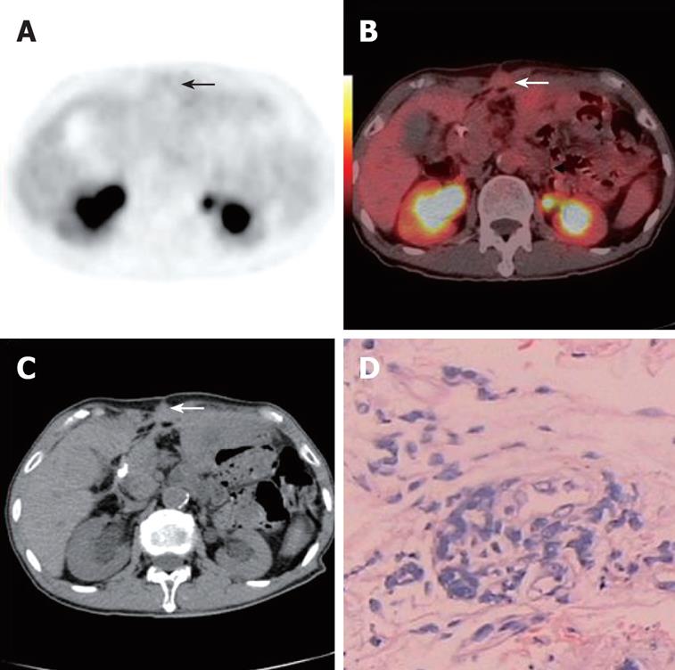
Figure 3 A 71-year-old asymptomatic man underwent PET/CT as part of routine post-operative surveillance after gastric cancer resection was performed 2.
5 years previously. Axial PET and PET/CT fusion images (arrows, A and B) showed no focal hypermetabolic activity in the abdominal wall. Axial contrast CT (white arrow, C) demonstrated local thickness in the abdominal wall. This was later verified as malignant by histopathological assessment of a CT guided core tissue biopsy (D).
- Citation: Sun L, Su XH, Guan YS, Pan WM, Luo ZM, Wei JH, Wu H. Clinical role of 18F-fluorodeoxyglucose positron emission tomography/computed tomography in post-operative follow up of gastric cancer: Initial results. World J Gastroenterol 2008; 14(29): 4627-4632
- URL: https://www.wjgnet.com/1007-9327/full/v14/i29/4627.htm
- DOI: https://dx.doi.org/10.3748/wjg.14.4627


