Copyright
©2008 The WJG Press and Baishideng.
World J Gastroenterol. Jul 28, 2008; 14(28): 4529-4534
Published online Jul 28, 2008. doi: 10.3748/wjg.14.4529
Published online Jul 28, 2008. doi: 10.3748/wjg.14.4529
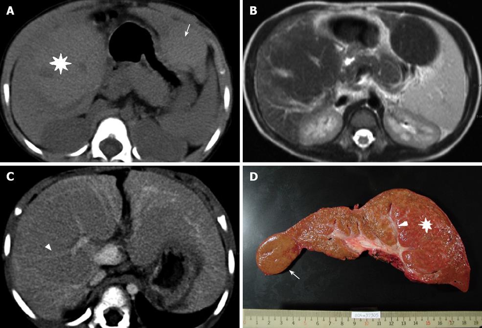
Figure 1 A 3-year-old BA girl with segments 5-8 (asterisk) and segment 2 (arrow) MRN.
A: The density of the MRN is slightly higher than that in the surrounding liver parenchyma during pre-enhanced phase of the CT; B: FSE/T2WI MRI shows a lower signal intensity in the MRN than in the surrounding liver; C: During portovenous phase of the CT, the tubular structure and splaying portal veins can be seen in the MRN (arrowhead); D: The explanted liver and intra-tumoral portal tract can be seen (arrowhead).
- Citation: Liang JL, Cheng YF, Concejero AM, Huang TL, Chen TY, Tsang LLC, Ou HY. Macro-regenerative nodules in biliary atresia: CT/MRI findings and their pathological relations. World J Gastroenterol 2008; 14(28): 4529-4534
- URL: https://www.wjgnet.com/1007-9327/full/v14/i28/4529.htm
- DOI: https://dx.doi.org/10.3748/wjg.14.4529


