Copyright
©2008 The WJG Press and Baishideng.
World J Gastroenterol. Jul 21, 2008; 14(27): 4389-4394
Published online Jul 21, 2008. doi: 10.3748/wjg.14.4389
Published online Jul 21, 2008. doi: 10.3748/wjg.14.4389
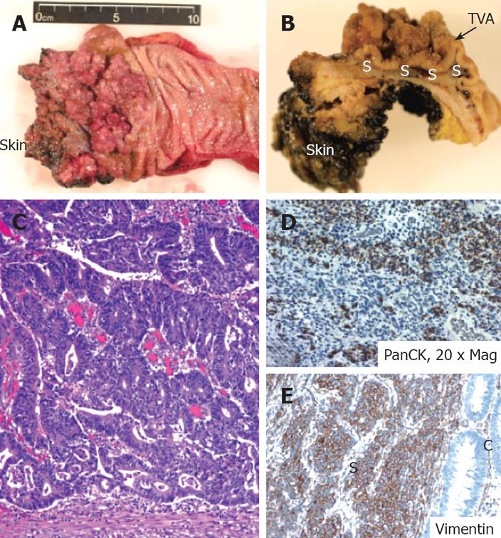
Figure 3 Gross appearance of the surgically-removed rectosigmoid mass (A and B).
A: The luminal side view demonstrates the proximity to the dentate line; anal skin is labeled for orientation; B: Upon sectioning the sample through the sagittal plane, the lateral view identifies the sarcomatoid smooth component (marked as S) underneath the velvety tubulovillous adenoma component (marked as TVA); C: Invasive adenocarcinoma with high nucleus/cytoplasm ratio within the deeper sections; D: Pancytokeratin was diffusely positive in both epithelial and mesenchymal components (× 20); E: Vimentin showed expression in the sarcomatous (S) component only, with no staining in the carcinoma (C) portion.
- Citation: Lee JK, Ghosh P, McWhorter V, Payne M, Olson R, Krinsky ML, Ramamoorthy S, Carethers JM. Evidence for colorectal sarcomatoid carcinoma arising from tubulovillous adenoma. World J Gastroenterol 2008; 14(27): 4389-4394
- URL: https://www.wjgnet.com/1007-9327/full/v14/i27/4389.htm
- DOI: https://dx.doi.org/10.3748/wjg.14.4389


