Copyright
©2008 The WJG Press and Baishideng.
World J Gastroenterol. Jul 21, 2008; 14(27): 4389-4394
Published online Jul 21, 2008. doi: 10.3748/wjg.14.4389
Published online Jul 21, 2008. doi: 10.3748/wjg.14.4389
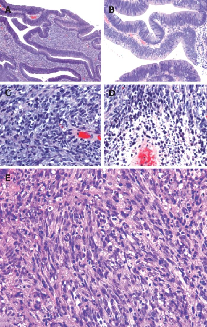
Figure 2 Histology of the rectal biopsy using HE stains.
A: Tubulovillous adenoma and underlying spindle cell tumor. The aggressive spindle cell lesion infiltrates directly underneath the adenoma (× 10); B: A higher magnification view of the adenomatous component (× 20). High-grade spindle cell lesion (× 40) showing: C: Cigar shaped nuclei, nuclear pleomorphism, a high mitotic rate; D: Tumor necrosis; E: Smooth muscle-like spindle sheets of cells in the sarcomatoid component (× 40).
- Citation: Lee JK, Ghosh P, McWhorter V, Payne M, Olson R, Krinsky ML, Ramamoorthy S, Carethers JM. Evidence for colorectal sarcomatoid carcinoma arising from tubulovillous adenoma. World J Gastroenterol 2008; 14(27): 4389-4394
- URL: https://www.wjgnet.com/1007-9327/full/v14/i27/4389.htm
- DOI: https://dx.doi.org/10.3748/wjg.14.4389


