Copyright
©2008 The WJG Press and Baishideng.
World J Gastroenterol. Jul 14, 2008; 14(26): 4253-4256
Published online Jul 14, 2008. doi: 10.3748/wjg.14.4253
Published online Jul 14, 2008. doi: 10.3748/wjg.14.4253
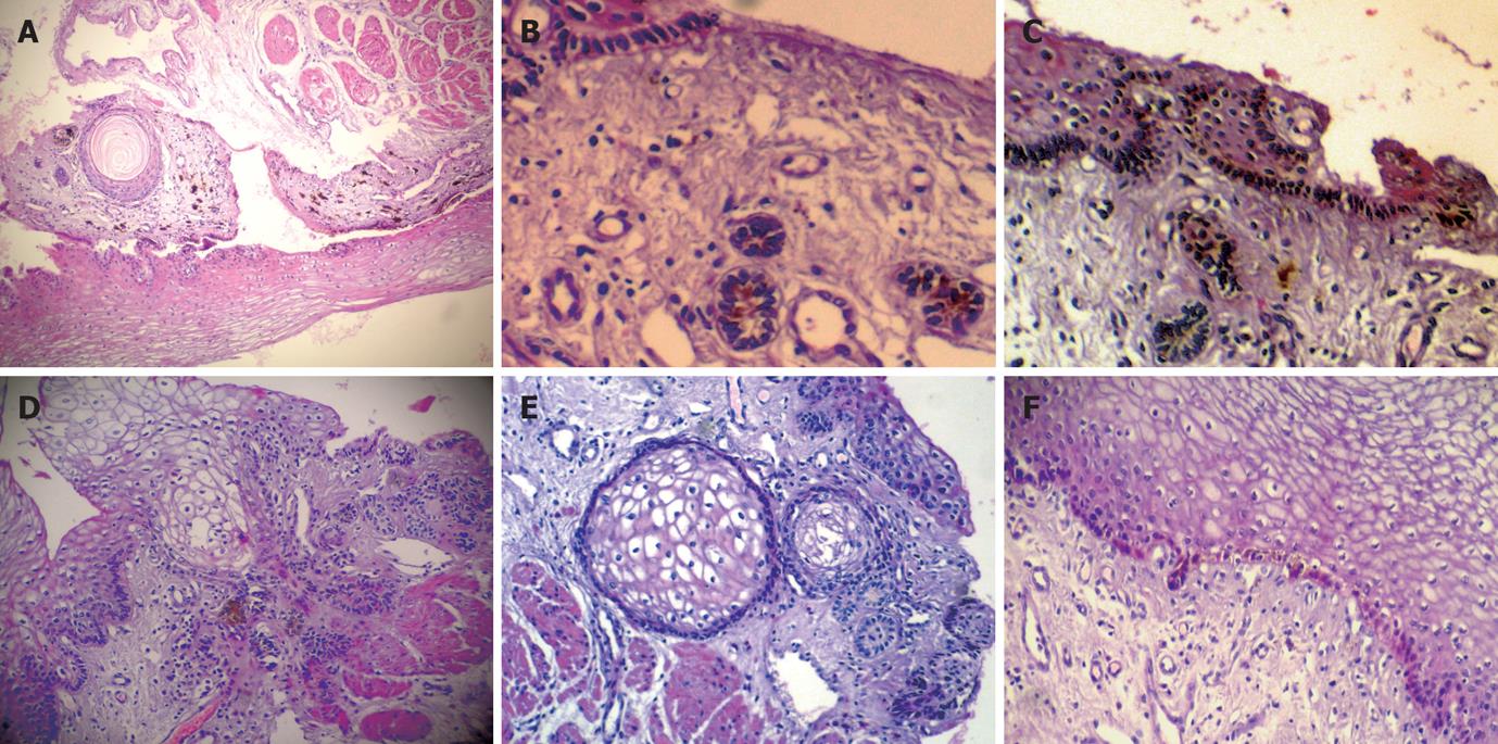
Figure 2 Focal distribution of melanocytes in keratinocytes of the basal esophageal mucosa layer, heavily pigmented, spindled or dendritic melanocytes/pigmentophages locate predominately in the superficial lamina propria, Moreover, there are several hair follicles and a horn cyst in the mucosa, with melanocytes in the hair follicle (HE, × 40) (A), and distribution of melanocytes in keratinocytes of the basal esophageal mucosa layer and hair follicles in the mucosal lamina propria with melanocytes (B,C) (B: HE, × 200; C: HE, × 40), cluster of hair follicles and horn cysts in the mucosa (D,E) (HE, × 40), and focal distribution of melanocytes in keratinocytes of the basal esophageal mucosa layer below the BSC (HE, × 40) (F).
- Citation: Wang DG, Li XG, Gao H, Sun XY, Zhou XQ. Coexistence of esophageal blue nevus, hair follicles and basaloid squamous carcinoma: A case report. World J Gastroenterol 2008; 14(26): 4253-4256
- URL: https://www.wjgnet.com/1007-9327/full/v14/i26/4253.htm
- DOI: https://dx.doi.org/10.3748/wjg.14.4253


