Copyright
©2008 The WJG Press and Baishideng.
World J Gastroenterol. Jul 14, 2008; 14(26): 4253-4256
Published online Jul 14, 2008. doi: 10.3748/wjg.14.4253
Published online Jul 14, 2008. doi: 10.3748/wjg.14.4253
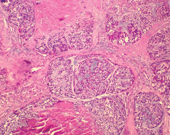
Figure 1 Arrangement of basaloid cells in the form of anastomosing trabeculae and microcystic structures.
The microcystic spaces contained basophilic mucoid matrix, mimicking an adenoid cystic carcinoma. The cells at the edges of basaloid islands tended to show peripheral nuclear palisading. Comedo necrosis was observed in the center of basaloid lobules (HE, × 40).
- Citation: Wang DG, Li XG, Gao H, Sun XY, Zhou XQ. Coexistence of esophageal blue nevus, hair follicles and basaloid squamous carcinoma: A case report. World J Gastroenterol 2008; 14(26): 4253-4256
- URL: https://www.wjgnet.com/1007-9327/full/v14/i26/4253.htm
- DOI: https://dx.doi.org/10.3748/wjg.14.4253


