Copyright
©2008 The WJG Press and Baishideng.
World J Gastroenterol. Jul 14, 2008; 14(26): 4249-4252
Published online Jul 14, 2008. doi: 10.3748/wjg.14.4249
Published online Jul 14, 2008. doi: 10.3748/wjg.14.4249
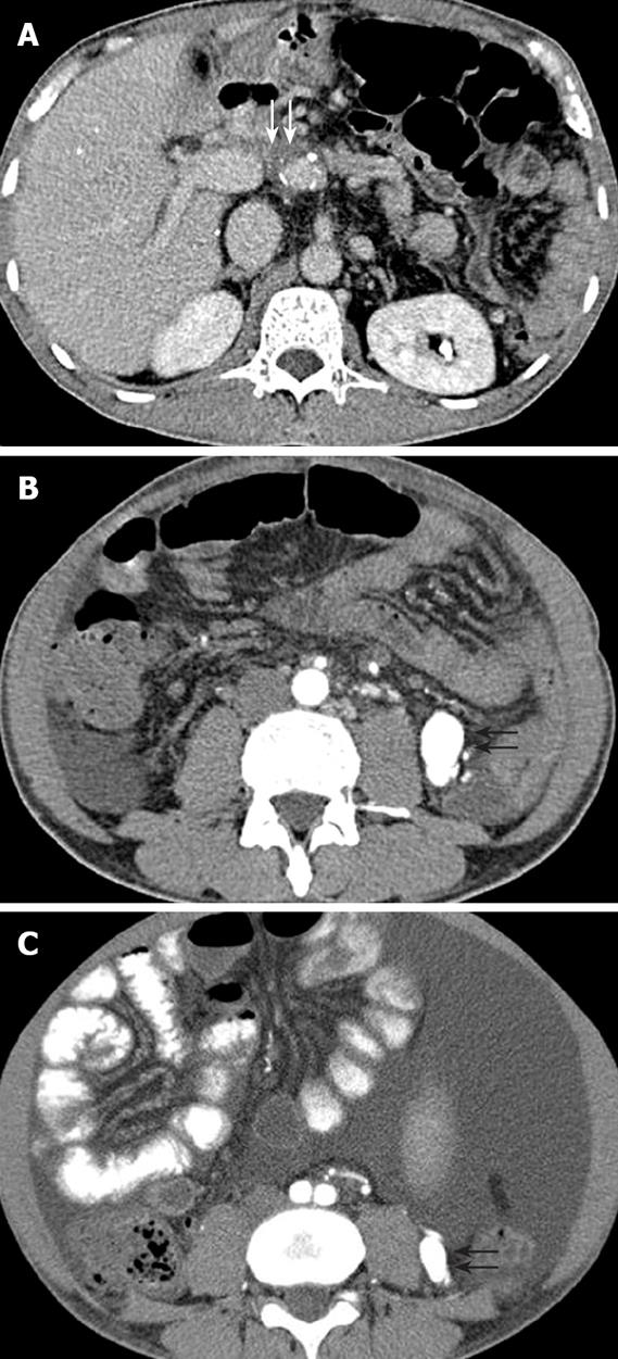
Figure 2 Contrast-enhanced CT of the abdomen showing portal vein stenosis (white arrows) (A), an approximately 30 mm × 18 mm contrast-enhancing vascular mass (black arrows) (B), and an approximately 10 mm × 8 mm enhancing vascular structure before transplantation (black arrows) (C).
- Citation: Kim IH, Kim DG, Kwak HS, Yu HC, Cho BH, Park HS. Ischemic colitis secondary to inferior mesenteric arteriovenous fistula and portal vein stenosis in a liver transplant recipient. World J Gastroenterol 2008; 14(26): 4249-4252
- URL: https://www.wjgnet.com/1007-9327/full/v14/i26/4249.htm
- DOI: https://dx.doi.org/10.3748/wjg.14.4249


