Copyright
©2008 The WJG Press and Baishideng.
World J Gastroenterol. Jul 14, 2008; 14(26): 4245-4248
Published online Jul 14, 2008. doi: 10.3748/wjg.14.4245
Published online Jul 14, 2008. doi: 10.3748/wjg.14.4245
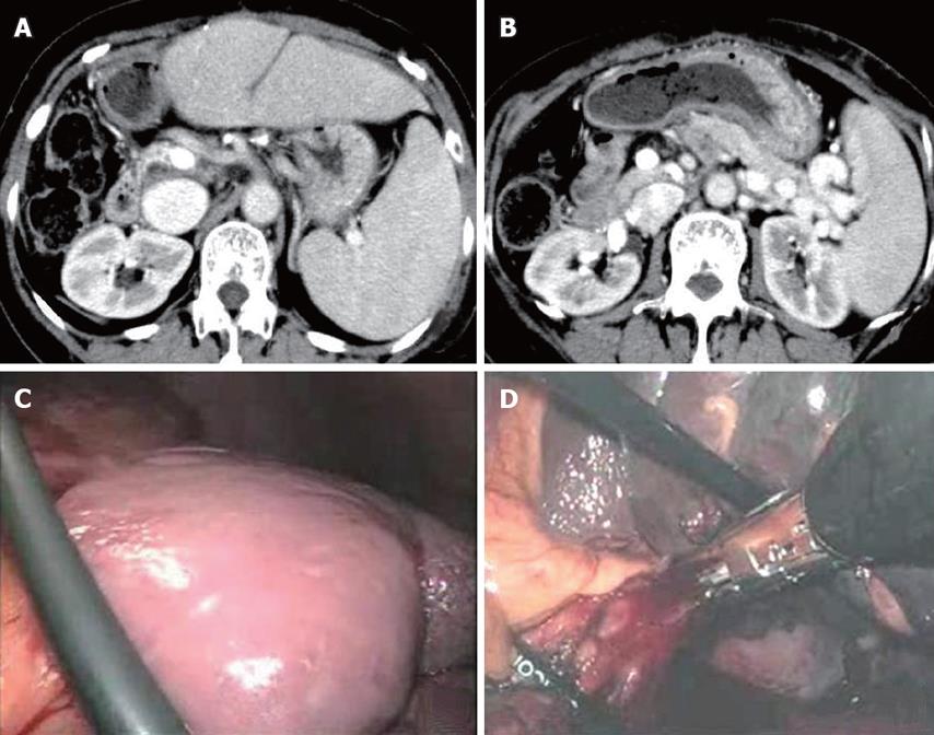
Figure 2 Abdominal enhanced CT showing splenomegaly with 140 mm in maximum diameter (A) and development of collateral veins around the splenic vein (B), no peritoneal adhesion to abdominal wall and around the spleen (C), and splenic hilum stapled with linear staplers (D).
- Citation: Kato H, Usui M, Azumi Y, Ohsawa I, Kishiwada M, Sakurai H, Tabata M, Isaji S. Successful laparoscopic splenectomy after living-donor liver transplantation for thrombocytopenia caused by antiviral therapy. World J Gastroenterol 2008; 14(26): 4245-4248
- URL: https://www.wjgnet.com/1007-9327/full/v14/i26/4245.htm
- DOI: https://dx.doi.org/10.3748/wjg.14.4245


