Copyright
©2008 The WJG Press and Baishideng.
World J Gastroenterol. Jul 14, 2008; 14(26): 4196-4203
Published online Jul 14, 2008. doi: 10.3748/wjg.14.4196
Published online Jul 14, 2008. doi: 10.3748/wjg.14.4196
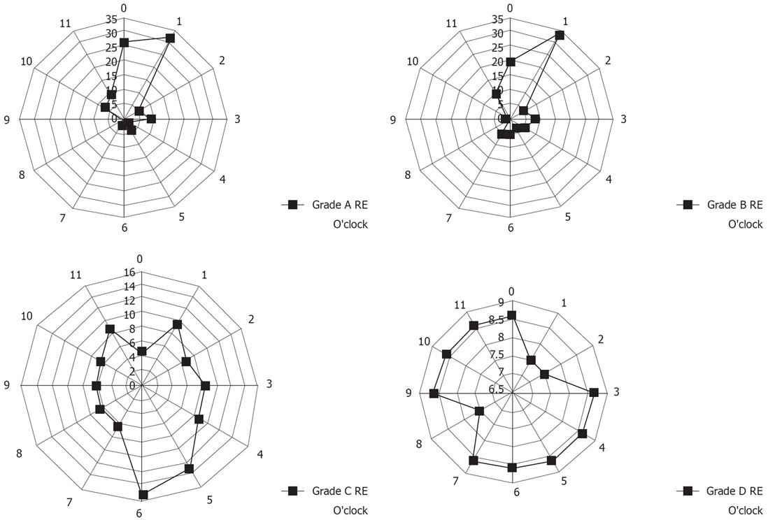
Figure 6 The circumferential location of esophageal mucosal breaks in subjects with grade A-D RE.
Data are shown in terms of clock face orientation. The numbers represent the percentage of lesions on each side of the esophageal wall relative to the total number of lesions. Patients with grade A and B esophagitis had longitudinal mucosal breaks mainly at the 12 o’clock and 1 o’clock wall of the lower esophagus, whereas patients with grade C and D esophagitis had transverse mucosal breaks mainly at the 4 o’clock to 6 o’clock wall of the lower esophagus. The circumferential distribution of esophageal mucosal breaks differed significantly among subjects with different grades of RE (χ2 test: P < 0.05, A vs B, A vs C, A vs D, B vs C, B vs D and C vs D).
- Citation: Yamagishi H, Koike T, Ohara S, Kobayashi S, Ariizumi K, Abe Y, Iijima K, Imatani A, Inomata Y, Kato K, Shibuya D, Aida S, Shimosegawa T. Tongue-like Barrett’s esophagus is associated with gastroesophageal reflux disease. World J Gastroenterol 2008; 14(26): 4196-4203
- URL: https://www.wjgnet.com/1007-9327/full/v14/i26/4196.htm
- DOI: https://dx.doi.org/10.3748/wjg.14.4196


